SAK BRACE

SAK BRACE specializes in medical orthopedic braces, rehabilitation supports, and customized OEM solutions for global distributors, clinics, and healthcare brands.

WhatsApp: 0086-13343185033 / 0086-13132404899 Email: info@sakbrace.com

Author name: admin

Category 1

PCL Brace vs OA Knee Brace — What’s the Real Difference?

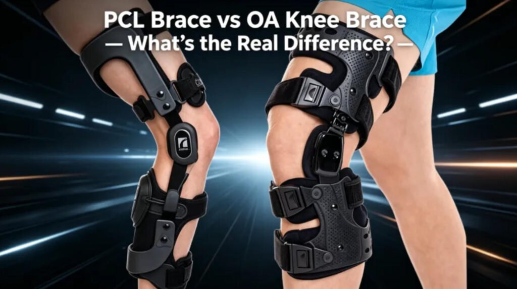
The Fundamental Difference Between Treatment Goals and Mechanisms of Action PCL (Posterior Cruciate Ligament) braces and OA (Osteoarthritis of the Knee) braces are two of the most common yet easily confused types of external fixation devices for the knee joint in orthopedic rehabilitation. Although both are functional knee orthotics, their design philosophies, biomechanical principles, and clinical applications are drastically different. The underlying difference stems from the fundamental problems they aim to solve—one addresses “instability,” while the other addresses “weight-bearing wear and tear.” PCL Braces: Precision Devices Against Bony Instability The posterior cruciate ligament (PCL) is one of the strongest ligaments in the knee joint, providing approximately 95% of the restraining force limiting posterior tibial slippage relative to the femur. When the PCL is damaged, patients experience “posterior tibial collapse” in a flexed knee position—the lower leg collapses posteriorly under gravity, leading to abnormal joint alignment. The core function of PCL braces is precisely to counteract this abnormal displacement. By placing a specialized support pad at the back of the lower leg, the brace actively pushes the tibia forward during knee flexion (especially near 90°), thereby reducing tension on the posterior PCL and creating a favorable biomechanical environment for ligament healing. Clinical guidelines from the NHS at Lancashire Teaching Hospitals in the UK explicitly state that the use of dynamic force braces can promote ligament healing over a period of time. Mechanically, commercially available professional PCL braces (such as the medi M.4s PCL dynamic and Össur Rebound PCL) typically employ a four-point multi-point stabilization system. Through a rigid frame and adjustable thrust device, they can precisely control the anterior displacement of the tibia. A clinical study published in Healthcare in 2026 showed that dynamic PCL braces significantly reduced posterior tibial displacement from an average of 7.1 mm to 2.68 mm under stress (p < 0.001), confirming their effective biomechanical control of posterior tibial displacement during the acute phase. From a clinical perspective, PCL braces primarily serve as a means of conservative treatment or postoperative rehabilitation after posterior cruciate ligament (PCL) injury, suitable for patients with acute or chronic PCL injuries. A typical usage protocol recommends wearing them for 24 hours daily (removing them only during showering) for approximately 16 weeks. However, it’s important to note that there is a significant gap in the evidence-based medicine regarding PCL braces. A comprehensive review published in Musculoskeletal Science and Practice in 2025, after systematically evaluating 1810 articles, indicated that the evidence for dynamic PCL braces is limited and inconsistent, lacking standardized treatment protocols, and there are no studies directly comparing the efficacy of different dynamic brace protocols. OA Braces: A Three-Point Lever System for Redistributing Joint Load The essence of knee osteoarthritis is degenerative wear and tear of the articular cartilage. For unicompartmental knee OA (most commonly medial compartment type), the core pathological issue is that the affected compartment bears excessive mechanical load. The design goal of OA braces is completely different from that of PCL braces—its task is not to “limit abnormal displacement,” but to “redistribute the load.” All knee osteoarthritis braces achieve load reduction through a three-point biomechanical principle: the femoral shell and calf shell form two stress points, while the dynamic strap system provides a third. Together, these three elements form a mechanical lever, transferring pressure from the affected compartment to the relatively healthy contralateral side. Taking Össur’s Unloader series as an example, its three-point lever system operates as follows: the brace applies a valgus force to the knee joint, actively opening the medial joint space and creating more space between the tibial plateau and femoral condyle, thereby reducing bone-on-bone contact and friction. The direct effect of this biomechanical intervention is pain relief and improved function, providing patients with a conservative treatment option beyond medication and surgery. In terms of clinical evidence, OA braces have a richer evidence-based foundation than PCL braces. A multicenter randomized controlled trial, PROP OA, published in the British Medical Journal (BMJ) in January 2026 (enrolling 466 patients aged 45 and older with knee OA), showed that adding compartment-specific knee braces to standard nonpharmacological treatment combined with adherence intervention resulted in a significant improvement in the knee outcome score (KOOS-5) at 6 months compared to the nonpharmacological treatment alone group (adjusted mean difference 3.39 points, 95% CI 0.96 to 5.82), with the most significant improvement in pain sub-scores (adjusted mean difference 6.13 points, effect size 0.39). Furthermore, a meta-analysis in 2025 indicated that among nonpharmacological interventions for knee OA, knee braces ranked highest in improving WOMAC pain, function, and stiffness scores, outperforming many other conservative treatments. However, this evidence is not without limitations—the BMJ study also found that the benefit of braces diminished over time, and the difference between groups was no longer statistically significant at 12 months. Researchers point out that declining adherence over time is a significant factor affecting treatment efficacy. Direct Comparison: Two Dramatically Different “Prescriptions” Comparing the two, the differences between PCL braces and OA braces can be summarized in the following core dimensions: Different Targets: PCL braces act directly on the ligament itself—reducing mechanical tension in the PCL through forward thrust, creating conditions for healing; OA braces, on the other hand, alter the overall biomechanical distribution of the knee joint through a three-point lever system—not acting on any ligament, but rather reducing contact pressure between the femoral condyle and tibial plateau. Different Failure Modes: PCL braces address “a sudden structural failure”—a ligament tear occurs at a specific moment, requiring external intervention to aid repair; OA braces address “a gradually degenerating system”—slow cartilage wear, requiring changes in the biomechanical environment to slow disease progression. Different Usage Modes PCL braces require 24-hour wear during the acute and rehabilitation phases (for several months); OA braces are recommended for use during weight-bearing activities (such as walking and climbing stairs), and can be removed during rest, with the wearing time gradually increased based on tolerance. The levels of evidence differ. OA braces have more high-quality randomized controlled trial evidence, especially in terms of clear data supporting

Category 1

ACL Brace vs PCL Brace: A Comparative Study of Two Types of Knee Ligament Braces

Abstract The anterior cruciate ligament (ACL) and posterior cruciate ligament (PCL) are core structures for maintaining sagittal stability of the knee joint, with opposite functional directions. Based on this biomechanical difference, ACL and PCL braces exhibit fundamentally opposing approaches in force line intervention, structural design, and clinical application strategies. This article systematically compares the two types of braces from four dimensions: anatomical basis, brace mechanics principles, clinical application scenarios, and current evidence-based practices, aiming to provide objective and professional reference for clinicians and rehabilitation practitioners. 1. Introduction Knee cruciate ligament injuries are common in sports medicine and orthopedics. ACL injuries are frequently seen in sports involving sudden stops and changes of direction (such as basketball, soccer, and skiing), while PCL injuries are often caused by “dashboard injuries” (direct posterior force on the proximal tibia) or falls while kneeling. Because the two types of ligaments are functionally antagonistic, the braces designed for their injuries follow drastically different biomechanical principles. However, in clinical practice, the two types of braces often appear similar, leading to frequent misuse or confusion. This can reduce rehabilitation effectiveness in mild cases and worsen ligament laxity or secondary joint instability in severe cases. Therefore, clearly distinguishing between ACL and PCL braces is a fundamental prerequisite for standardized conservative treatment and postoperative rehabilitation. 2. Anatomical and Biomechanical Basis The ACL originates from the medial surface of the lateral femoral condyle, runs anteromedially, and inserts anterior to the intercondylar eminence of the tibia. Its main function is to limit excessive anterior displacement of the tibia relative to the femur, while also assisting in controlling knee joint rotational stability. The PCL originates from the lateral surface of the medial femoral condyle, runs posterolaterally, and inserts posterior to the intercondylar eminence of the tibia. It is the primary structure preventing posterior displacement of the tibia. The PCL has approximately twice the cross-sectional area of the ACL and a higher ultimate tensile strength, thus resulting in a relatively lower injury rate. During knee flexion and extension, the ACL bears the main tension under anterior shear force, while the PCL is stretched under posterior shear force. When a brace attempts to protect a ligament, it must apply an external restraint force opposite to the direction of the pathogenic force: ACL braces need to counteract anterior tibial displacement, while PCL braces need to counteract posterior tibial displacement. This mechanical opposition forms the root of all design differences between the two types of braces. 3. Core Comparison of Brace Design Principles 3.1 Direction of Restraint Force The core load-bearing direction of ACL braces is posterior. That is, the brace mechanically prevents the tibia from sliding forward by applying posterior pressure to the anterior part of the tibia. To achieve this, most functional ACL braces are equipped with a rigid beam or padding on the anterior side, which, together with a hinge and strap system, maintains anterior obstruction during knee flexion and extension. The core load-bearing direction of PCL braces is completely opposite—anterior. PCL braces actively push the tibia forward by applying a continuous anterior thrust to the posterior proximal end of the tibia, thereby reducing the tension on the PCL. This need spurred the design of the “dynamic PCL brace”: featuring a posterior elastic pad or airbag and an adjustable spring loading system to maintain a constant forward corrective force throughout the joint’s flexion and extension range. 3.2 Structural Feature Comparison It is worth noting that static ACL braces (postoperative immobilization type) typically fix the knee joint in full extension or slight flexion to reduce graft stress; while postoperative PCL braces often use posterior support in extension to prevent posterior tibial subsidence due to gravity. 4. Clinical Indications and Application Strategies 4.1 Applicable Scenarios for ACL Braces ACL injuries can be classified into Grade I (minor stretching), Grade II (partial tear), and Grade III (complete rupture). For patients with Grade I-II injuries or a strong desire for conservative treatment, functional ACL braces are used to: limit abnormal forward translation and rotational movements; assist proprioceptive reconstruction; reduce the risk of re-injury during movement (the strength of evidence is controversial). After ACL reconstruction, the brace is usually worn for 4-6 weeks, initially limiting the knee flexion angle (e.g., 0°-90°), gradually loosening it. The recently developed “cross brace approach,” which uses a 90° knee flexion position for long-term fixation, explores a new non-surgical treatment path for high-grade tears, but it is still in the research stage. 4.2 Applicable Scenarios for PCL Braces PCL injuries are also graded according to laxity. Conservative treatment is recommended for Grade I-II acute injuries. Dynamic PCL braces can effectively eliminate posterior subsidence, reduce pain, and promote ligament alignment and healing. Treatment of grade III isolated PCL injuries remains controversial, but for patients not opting for surgery, dynamic bracing combined with early functional rehabilitation is one of the mainstream approaches. Postoperative management of PCL reconstruction is more stringent: A brace with posterior support in full extension is typically used, with partial weight-bearing (up to 20 kg) for the first 6 weeks post-surgery, and knee flexion limited to 90°. Dynamic braces (such as PCL Jack braces) provide continuous anterior force post-surgery, helping to protect the reconstructed ligament. 4.3 Absolutely Prohibited: Mixed Use of Braces Using an ACL brace for a PCL injury will result in complete inability to control posterior subduction due to insufficient anterior blockage or lack of posterior thrust; conversely, using a PCL brace for an ACL injury will increase tension on the ACL or graft due to continuous anterior thrust, causing secondary injury. There are clearly reported cases of ACL reconstruction graft laxity due to misuse of PCL braces. 5. Evidence and Existing Controversies 5.1 Evidence Dilemmas Regarding ACL Braces Multiple systematic reviews show that functional ACL braces can reduce ACL strain under forward shear loads under laboratory conditions, but their control over rotational stability during dynamic movements (including lateral shearing and sudden stops) is inconsistent. Randomized controlled trials have contradictory conclusions: some studies support that braces reduce the risk of re-tears

Uncategorized

How PCL brace improves posterior tibial control: A typical case analysis

1. What problems did the patient face? A 35-year-old male, an avid sports enthusiast, suffered a grade II tear of the right posterior cruciate ligament (PCL) during a soccer match due to a direct frontal impact while the knee was flexed. The patient complained of posterior knee pain and swelling, which worsened, especially when climbing stairs, squatting, and walking for extended periods. Physical examination revealed a positive posterior drawer test (approximately 8 mm posterior tibial displacement) and significant posterior tibial tuberosity subsidence. Gait analysis showed a compensatory pattern of “knee flexion and forward lean” during gait—that is, consciously contracting the quadriceps and reducing knee flexion during the standing phase to prevent abnormal posterior tibial displacement due to gravity and hamstring traction. The patient’s core functional impairment was not simply pain or limited mobility, but rather loss of posterior tibial stability. In cases of PCL dysfunction, the tibia abnormally sinks posteriorly and downward under weight-bearing, altering the knee’s flexion-extension center. This leads to increased pressure on the patellofemoral joint, compensatory tension in the medial collateral ligament, and accelerated degeneration of the posterior joint structures. 2. What are the biomechanical causes? The PCL is the most important posterior stabilizing structure of the knee joint. Its core function is to limit posterior displacement of the tibia relative to the femur. In a normal knee joint, the PCL bears approximately 85%-95% of the anti-posterior displacement load within a 30° to 120° flexion range. When the PCL is injured, the biomechanical chain is disrupted: Gravity: When squatting or descending stairs, gravity pushes the tibial plateau posteriorly through the femoral condyle. Without PCL restraint, the tibia sinks abnormally posteriorly. Muscle imbalance: The hamstring muscles (semitendinosus, semimembranosus, biceps femoris) cross the posterior aspect of the knee joint, and their contraction actively pulls the tibia posteriorly. In PCL defects, this physiological tension transforms into a harmful displacement force. Lack of proprioceptive feedback: The PCL contains abundant mechanoreceptors; after injury, joint positional sense decreases, and patients cannot perceive whether the tibia is in a neutral position. The result: With every weight-bearing movement, the tibia repeatedly and abnormally shifts backward. This small but repetitive displacement gradually stretches the posterior joint capsule, irritates the synovium, and wears down the posterior horn of the meniscus, leading to secondary joint instability. 3. Mechanism of Action of PCL Braces To address the aforementioned biomechanical deficiencies, PCL-specific braces (such as posterior support knee braces) are designed to actively resist posterior tibial displacement, rather than simply providing medial and lateral stability like ordinary hinged braces. The PCL brace worn by this patient has three key structures: Posterior tibial shelf: Located approximately 2 cm below the popliteal fossa, it directly pushes forward against the posterior cortex of the proximal tibia. When the tibia attempts to move backward, the shelf provides an anterior reaction force. Adjustable anterior pull strap: This strap wraps around the front of the lower leg from the rear frame of the brace. When tightened, it generates a continuous forward force, similar to “pushing the tibia forward from behind.” Quadriceps-assisted hinge: The brace provides extension assistance at the end of knee extension, encouraging the patient to use their quadriceps to control the knee joint, while allowing the hamstrings to relax relatively. Through these three mechanisms, the brace achieves functional anterior restraint. Even in a relaxed standing position, the posterior tibial subsidence sign largely disappeared after wearing the brace. 4. Clinical Benefit Assessment This patient wore the PCL brace for 8 hours daily (during daytime activities) in conjunction with rehabilitation training. A follow-up examination after 4 weeks showed: Static stability: Posterior drawer test showed posterior tibial displacement reduced from 8mm to 3mm (close to the normal range). Dynamic control: The “sudden sinking” sensation during squatting disappeared; the gait returned to normal when climbing stairs, and the compensatory pattern disappeared. Pain score: VAS decreased from 6/10 to 2/10, primarily related to reduced joint capsule tension. Functional score: IKDC subjective knee score improved from 52 to 78. Muscle activation pattern: Surface electromyography showed a decrease of approximately 30% in the hamstring/quadriceps co-activation ratio, indicating that excessive compensatory contraction is no longer needed. It is worth noting that the brace does not completely replace PCL function, but rather provides a biomechanical “crutch”—protecting the joint and allowing patients to begin early functional training before ligament healing or reconstruction surgery. Literature indicates that with conservative treatment using a PCL brace for grade II PCL injuries, approximately 70% of patients can regain near-normal posterior stability after 3 months. 5. Summary The core issue of PCL injuries is abnormal posterior displacement of the tibia during weight-bearing, rather than simply pain or decreased range of motion. This displacement can lead to secondary joint damage. PCL-specific braces provide continuous anti-posterior restraint through posterior pads and anterior tension bands, directly counteracting gravity and adverse hamstring forces. Clear clinical benefits include improved static and dynamic posterior tibial control, pain reduction, gait restoration, and reduced abnormal muscle compensation. However, braces are not a panacea: they must be combined with quadriceps strengthening and neuromuscular retraining; grade II or higher tears or those with multiple ligament injuries still require evaluation for surgical indications. Brace designs guided by biomechanical principles are better able to address the specific problems following PCL loss than general-purpose knee braces. For patients who are unwilling or temporarily unable to undergo surgery, a PCL brace is currently one of the most well-supported conservative interventions.

Uncategorized

Understanding PCL Injury Grades: From Grade 1 to Grade 3

The posterior cruciate ligament (PCL) is one of the thickest and strongest ligaments in the knee joint, located in the center of the knee. Its main function is to limit the posterior displacement and external rotation of the tibia relative to the femur. PCL injuries account for approximately 1% to 47% of acute knee ligament injuries, mostly occurring in severe collisions such as car accidents or contact sports. Clinically, PCL injuries are classified into three grades—Grade I, Grade II, and Grade III—based on the posterior drawer test of the knee joint. This grading system is primarily based on the posterior displacement of the tibia in the posterior drawer test, combined with pathological changes in the ligament and joint stability for a comprehensive assessment. Grade 1 Injury: Mild – Microscopic Tear, Remaining Stable Grade 1 injuries are mild, characterized by a small tear or microscopic tear of the ligament fibers. Pathologically, the ligament fibers are stretched but the overall structure remains intact, or only a very few fiber bundles are ruptured. In the posterior drawer test, the posterior displacement of the tibia relative to the femur is less than 5 mm. At this stage, the tibial plateau remains anterior to the femoral condyle. Clinically, patients with Grade 1 injuries typically do not experience significant joint instability; some may even be unaware of the injury or have no pain. These injuries are usually treated conservatively, including rest, ice application, compression bandages, and limb elevation. Grade 2 Injury: Moderate – Partial Tear, Impaired Stability Grade 2 injuries are moderate, characterized by the rupture of more ligament fibers, representing a partial tear of the ligaments. The integrity of the ligaments is significantly compromised, but not completely severed. In the posterior drawer test, the tibia shifts posteriorly by 5–10 mm. At this stage, the tibial plateau is essentially level with the femoral condyle. Clinically, patients with Grade 2 injuries experience mild to moderate joint instability, and may feel weakness in the knee joint when standing, walking, or undergoing physical examination. Conservative treatment is usually the first consideration for these injuries, and with bracing and systematic rehabilitation training, most patients achieve good results. Grade 3 Injury: Severe – Complete Tear, Significant Instability A Grade 3 injury is a severe injury, characterized by a complete rupture of the ligament. In the posterior drawer test, the tibia shifts posteriorly by more than 10 mm, with the tibial plateau regressing posterior to the femoral condyle. Significant knee instability is observed at this stage. It is important to note that Grade 3 injuries are often not isolated and are frequently accompanied by injuries to other important knee structures, such as posterolateral horn injuries, medial collateral ligament injuries, or anterior cruciate ligament injuries. Treatment strategies for this type of injury are somewhat controversial. Conservative treatment may be attempted for elderly patients or those with low activity levels; while for younger patients with high activity levels or those for whom conservative treatment is ineffective, arthroscopic PCL reconstruction surgery should be considered. Surgical indications are more clearly defined in complex cases such as multiple ligament injuries or meniscus root avulsion. Diagnosis and Assessment The grading and diagnosis of PCL injuries rely on standardized physical examination and imaging evaluation. The posterior drawer test is the most crucial physical examination method for diagnosis and grading. MRI has a sensitivity of 95%–100% for diagnosing PCL injuries, clearly showing ligament continuity and signal changes, and is an important imaging tool for assessing the degree of injury. Stress X-rays can provide quantitative evidence for grading by objectively measuring the difference in posterior tibial displacement between the two knees. Prognosis and Rehabilitation The PCL has a certain self-healing ability, especially the middle and distal third of the solid portion, which has a rich blood supply and strong healing potential after rupture. Therefore, for acute grade 1 and 2 PCL injuries, conservative treatment should be actively pursued. Studies have shown that patients with isolated grade 2 or 3 PCL injuries who receive conservative treatment can return to sports on average after 16 weeks. For patients undergoing surgical treatment, systematic rehabilitation training is required postoperatively, and long-term follow-up is recommended to assess knee joint function recovery and the occurrence of complications. Overall, the grade 1–3 PCL injury grading system provides clinicians with a systematic framework from pathological changes and the degree of joint instability to the selection of treatment strategies. Accurate injury grading is a prerequisite for developing individualized treatment plans, predicting prognosis, and guiding rehabilitation. For the general public, understanding this classification system helps in seeking timely medical attention after a knee injury, accurately describing symptoms, and communicating effectively with doctors.

Category 1

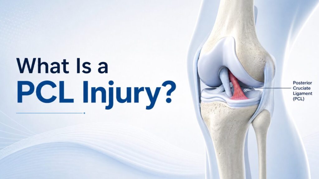
What Is a PCL Injury? The Often-Overlooked Ligament Behind Knee Stability

I. What is the Posterior Cruciate Ligament (PCL)? The posterior cruciate ligament (PCL) is the largest and strongest ligament in the knee joint. Together with the anterior cruciate ligament (ACL), it forms the core cruciate ligament system of the knee joint, crossing in an “X” shape inside the knee. The PCL originates from the lateral surface of the medial femoral condyle, running obliquely posterolaterally, and inserts posterior to the intercondylar eminence of the tibia. The PCL consists of two bundles of fibers with different functions: the larger anterolateral bundle (accounting for approximately 95% of the ligament volume) and the smaller posteromedial bundle (accounting for approximately 5%). The anterolateral bundle is most taut when the knee is flexed at approximately 70°, while the posteromedial bundle plays a major role near extension. This intricate fiber arrangement allows the PCL to continuously provide stable posterior support at various angles of the knee joint. The peristaltic ligament (PCL) is the primary structure limiting posterior displacement of the tibia relative to the femur, especially when the knee is flexed beyond 30°, providing approximately 95% of the resistance to posterior tibial displacement. Furthermore, the PCL provides rotational stability when the knee is flexed beyond 90°. In short, the PCL is the “first line of defense” for posterior knee stability. II. Mechanisms of PCL Injuries PCL injuries are not the most common type of knee ligament injury—accounting for about 3% of outpatient knee injuries—but can reach as high as 38% in acute traumatic knee hematoma. The PCL is the least likely knee ligament to be injured during exercise; the vast majority of PCL injuries are accompanied by damage to other knee ligaments or structures, and isolated PCL injuries are relatively rare. The classic mechanism of a PCL injury is that an external force acts directly on the anterior aspect of the tibia, subjecting the tibia to a posterior impact force. The most common scenarios include: Car accident injuries: also known as “dashboard injuries”—when a passenger’s bent knee strikes the car’s dashboard during sudden braking or a collision, the upper tibia receives a backward force, resulting in a PCL tear. Sports injuries: athletes fall forward with their knees bent, causing the tibial tuberosity to directly impact the ground. This is common in sports such as American football, rugby, basketball, and skiing. Additionally, sudden deceleration or change of direction while running, with the foot fixed to the ground, can also cause PCL injuries without direct impact. Hyperextension injuries: when the knee joint is forcibly and passively extended, the PCL may also suffer traction injuries. III. Injury Grading PCL injuries are generally classified into three grades based on severity: Grade I (Mild): The ligament has only microscopic tears; the ligament is stretched but the overall structure remains intact, and knee joint stability is largely unaffected. Grade II (Moderate): Partial ligament tear; the knee joint exhibits mild to moderate laxity and instability. Under stress testing, the posterior displacement of the tibia is 5–10 mm. Grade III (Severe): Complete ligament rupture or avulsion of the ligament from its bony insertion, resulting in significant knee instability. The tibia shifts posteriorly by more than 10 mm. Grade III injuries typically require substantial external force and are often accompanied by damage to the ACL, collateral ligaments, or other knee joint structures. IV. Clinical Manifestations and Diagnosis The symptoms of PCL injuries are sometimes atypical, which can lead to missed diagnoses. Common manifestations include: mild knee swelling, posterior knee pain (worsened by kneeling), and anterior knee pain during running or deceleration (which may appear one to two weeks after the injury). Many athletes can continue competing even after an injury, only realizing later that their knee is “not right.” In physical examination, the posterior drawer test is the most sensitive and specific method for diagnosing PCL injuries. The physician flexes the patient’s knee to 90° and pushes the tibia posteriorly; if the tibia shifts significantly posteriorly compared to the contralateral side, it suggests a PCL injury. In terms of imaging, MRI is the gold standard for diagnosing PCL injuries, clearly showing the location and extent of ligament damage. Stress radiographs can objectively assess the degree of posterior tibial displacement and are of significant value in determining the need for surgery. Plain radiographs are valuable for diagnosing avulsion fractures at the ligament insertion point. V. Treatment Strategies The choice of treatment plan for PCL injuries requires comprehensive consideration of factors such as injury grade, presence of other injuries, patient age, and exercise needs. Conservative treatment is suitable for grade I and II isolated PCL injuries. Due to the rich blood supply to the PCL and its strong self-healing ability, conservative treatment often achieves good results. The core components of conservative treatment include: early bracing and immobilization (usually around 6 weeks), control of swelling and pain, and systematic rehabilitation training. The focus of rehabilitation training is quadriceps strengthening exercises while delaying hamstring activation to avoid posterior tibial pull from hamstring contraction during early training, which could worsen ligament damage. Studies show that with physical therapy-guided exercise and bracing intervention, patient-reported outcomes can significantly improve during long-term follow-up. Surgical treatment is suitable for patients with grade III complete injuries, multiple ligament injuries, or significant instability after conservative treatment. Currently, the mainstream surgical procedure is arthroscopic PCL reconstruction, which can be divided into single-bundle reconstruction and double-bundle reconstruction based on the number of reconstructed bundles. Studies have shown that anatomical double-bundle reconstruction can better restore the original ligament coverage area and normal knee joint kinematics. For isolated PCL injuries with posterior tibial displacement ≥10mm, surgical reconstruction is a clear indication. VI. Prognosis and Long-Term Impacts If PCL injuries are not properly treated, they can lead to long-term adverse consequences. PCL dysfunction alters knee joint kinematics, leading to increased load on the medial compartment and patellofemoral joint, thereby accelerating articular cartilage degeneration. Studies have reported that the incidence of medial femoral condyle cartilage degeneration can reach 77.8% five years after a PCL defect. However, the treatment of PCL injuries remains

Uncategorized

From Rehabilitation to Prevention: An Objective Examination of the Value of Braces in Sports Injuries

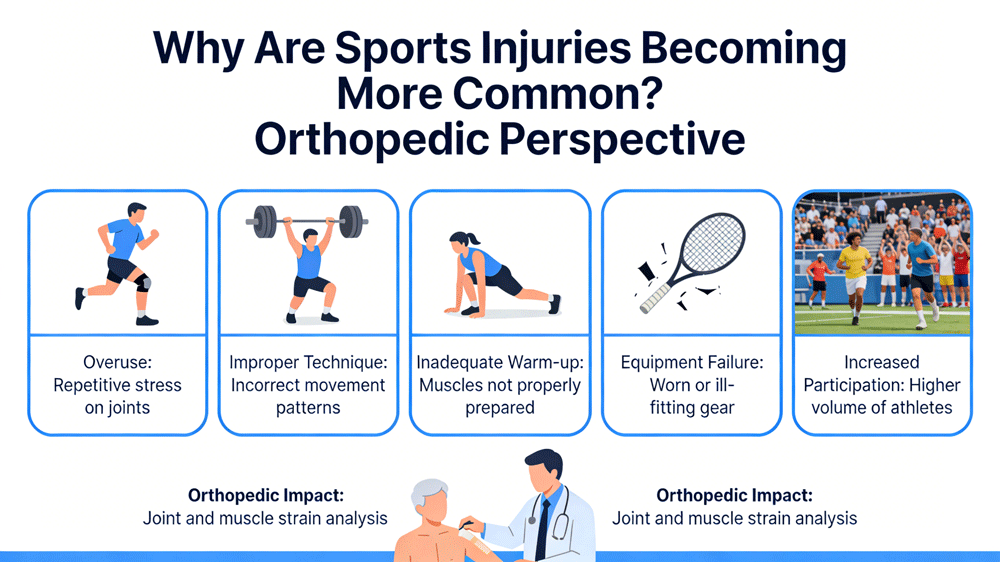
In today’s nationwide fitness boom, sports injuries have become an almost inevitable consequence of this enthusiasm, whether for marathon runners, weightlifting enthusiasts, or amateur athletes. As a practitioner/enthusiast focusing on sports rehabilitation, I’ve recently found in discussions with colleagues that opinions on braces are often polarized: some see them as a lifeline after injury, while others consider them a restrictive burden. Today, I want to discuss the topic of “Common Sports Injuries and Braces” objectively, without subjective bias. I. The Core Role of Braces: Not Healing, But Managing First, we need to clarify a concept: braces themselves do not possess healing capabilities; they cannot heal torn ligaments or eliminate inflammation. Their core function is biomechanical intervention and management. At different stages of sports injuries, braces play three distinct roles: 1. Immobilization Phase: After the acute phase of the injury or post-operatively, braces restrict movement in specific directions, providing a tension-free and stable environment for soft tissue healing. 2. Functional Phase: As tissues begin to heal, a balance needs to be struck between protection and mobility. Functional braces allow joints to move within a safe range, preventing muscle atrophy and joint stiffness. 3. Prophylactic Phase: This is the most controversial yet most widely used area, where braces are used by healthy individuals or those with prior injuries to prevent injury or recurrence during high-risk sports. II. Objective Analysis of Common Injuries and Corresponding Braces Let’s look at some common examples in sports scenarios: 1. Knee Joint: Anterior Cruciate Ligament (ACL) Injury Common Scenarios: Sudden stops and changes of direction in basketball and soccer. Brace Application: Hinged knee braces are standard in post-operative rehabilitation. It controls the extension angle, preventing overextension that strains reconstructed ligaments. Objective Perspective: For ACL injuries, the academic community generally agrees that postoperative bracing is part of the rehabilitation process. However, the evidence for the effectiveness of prophylactic braces in preventing first-time injuries is insufficient. It may give the wearer a false sense of invulnerability, leading to more dangerous movements. 2. Ankle: Recurrent Sprains Common Scenarios: Running, badminton, outdoor hiking. Brace Application: Semi-rigid or soft ankle braces. Objective Perspective: Compared to the knee, ankle braces have more robust evidence for prevention. For individuals with a history of multiple sprains, wearing a brace during high-intensity activities (such as contact sports and trail running) can significantly reduce the risk of re-sprains. However, it should not replace proprioceptive training—the brain’s control over ankle position is the ultimate “ankle protection.” 3. Wrist: Scaphoid Fracture / Triangular Fibrocartilage Complex Injury Common Scenarios: Falling and landing on the palm (skiing, skateboarding), gymnastics. Brace Application: Wrist immobilization brace. Objective Perspective: Wrist braces are often synonymous with “absolute immobilization.” Due to the unique blood supply to the scaphoid bone, healing is slow, requiring high compliance with braces. Many enthusiasts remove the brace themselves once the pain subsides, often leading to nonunion—a very serious misconception. 4. Lower Back: Spondylolysis / Lumbar Disc Herniation Common Scenarios: Weightlifting, golf, sudden physical activity after prolonged sitting at a desk. Brace Application: Rigid or soft lumbar support. Objective Perspective: The core function of a lumbar brace is to increase intra-abdominal pressure and distribute the load on the lumbar spine. It is an excellent aid during extreme weight-bearing activities (such as heavy squats). However, prolonged reliance on lumbar support in daily life can lead to core muscle atrophy and weakening, making the lumbar spine more vulnerable. III. Three Objective Suggestions for Sports Enthusiasts Based on the above observations, I have three preliminary thoughts for those considering or currently using braces: 1. Understand the “Expiration Date” of Braces Braces are “crutches” in the rehabilitation process, not permanent “prosthetics.” Except for a few athletes with special needs, most people’s ultimate goal should be to get rid of braces and regain joint control through rehabilitation training. 2. Beware of “Brace Dependence” and “Brace Illusion” On the one hand, over-reliance on braces can weaken proprioception; on the other hand, the sense of security provided by wearing braces may induce actions beyond the current joint’s capacity, causing more insidious injuries. 3. The Importance of Personalized Customization There is a wide variety of sports braces on the market. For simple sprains, a general-purpose brace may suffice; however, for complex ligament injuries or post-operative rehabilitation, a brace custom-made by a rehabilitation physician or orthotist provides more precise pressure distribution and range of motion control, far exceeding the value of a regular “protective gear.” In Conclusion Sports injury rehabilitation is an art of “balance.” A brace is a precise ruler in this artistic creation, but it cannot draw the blueprint for health. True freedom of movement comes from strong muscle strength, sensitive neural control, and a scientific training plan. A brace is merely a reliable and rational partner on our journey back to the track. Interactive Topic: Have you ever been injured while exercising? What was your experience with the brace or protective gear you used? Feel free to share your experiences and opinions in the comments section, and let’s exchange tips on avoiding common pitfalls.#SportsRehabilitation #SportsInjury #HealthEducation #PhysicalTherapy #WorkplaceHealth

Uncategorized

Medical Braces vs. Sports Protective Gear: Don’t Confuse Them – Choose the Right One for True Protection

In rehabilitation clinics or sports fields, I often encounter two types of people: one type is patients who come in wearing sports knee braces after ligament injuries, complaining, “Why is my knee still wobbly even with the brace?”; the other type is novice runners who wear heavy medical hinge braces to races, only to find they can’t run and have their skin chafed. This reflects a common misconception: confusing medical braces with sports protective gear. Although they look similar and are both worn on the limbs, they belong to two completely different technological paths in terms of design logic, clinical purpose, and usage scenarios. Today, we will break down the essential differences between the two from a professional, objective, and realistic perspective. Core Difference: Clinical Treatment vs. Sports Protection Medical braces, often referred to as “orthoses” in professional terminology, fall under the category of rehabilitation engineering. Their core logic is “replacement and reconstruction.” When the musculoskeletal system loses stability due to trauma, surgery, or nerve damage, medical braces temporarily replace the function of damaged ligaments or bones through external rigid structures. For example, adjustable hinged braces worn by patients after anterior cruciate ligament reconstruction aim to lock the knee joint in extension to prevent hyperextension and gradually unlock the range of motion according to the healing progress. In clinical applications at the China Rehabilitation Research Center, walking orthoses for spinal cord injury patients (such as ARGO) even have assistive functions, helping paraplegic patients stand again. These braces typically feature metal or high-strength plastic supports and angle adjustment chucks, with biomechanical stability as the primary design consideration, rather than comfort or portability. Sports protective gear falls under the category of sports protection. Its core logic is “prevention and assistance.” During exercise, protective gear enhances proprioception and reduces soft tissue concussion by applying pressure, insulation, and appropriate restriction, thereby reducing the risk of injury. Take, for example, the elastic knee brace commonly used by basketball enthusiasts. It primarily provides even pressure, promotes blood circulation, and reminds the wearer to “be careful here,” preventing the joint from deviating excessively from its physiological trajectory. For runners without injuries, sports braces serve more as a psychological safety net and provide mild physical protection. They are typically made of elastic fibers, knitted fabrics, and silicone anti-slip strips, emphasizing breathability, fit, and flexibility. By Cleo Hsieh The Dangers of Scenario Substitution What are the consequences of incorrectly substituting the two? Case A: Using Sports Braces Instead of Medical Braces A patient undergoing conservative treatment for an anterior cruciate ligament injury purchased a sports knee brace costing several hundred yuan to save money or for convenience. As a result, when walking and turning, because the sports knee brace only provides circumferential pressure and lacks anterior-posterior rigid support, the knee joint shifted instantly, leading to a secondary meniscus tear. Professional medical braces, such as post-operative knee braces, precisely control the flexion and extension range of the knee joint through bilateral hinges and a rigid frame, something ordinary elastic knee braces cannot achieve. Case B: Using Medical Braces for Daily Activities A young man, after his fracture had healed, continued to wear a heavy, low-temperature thermoplastic brace to play badminton. He found that not only could he not run, but because the brace completely restricted ankle movement, he couldn’t cushion his landings, causing stress to be directly transmitted to his lower leg, leading to stress periostitis. Medical braces are designed to provide absolute immobility in the early stages of rehabilitation; over-reliance can lead to muscle atrophy and decreased proprioception, actually increasing the risk of re-injury. How to Choose Scientifically? Consider Three Dimensions So, faced with a dazzling array of products on the shelves, how should professionals or consumers choose? It is recommended to consider the following three dimensions: Consider the Stage of Injury (Acute Phase vs. Recovery Phase vs. Prevention Phase) Post-operative/Acute Injury Phase (0-6 weeks): Choose a medical brace unconditionally. For example, after ACL surgery, an adjustable knee brace should be used, and the angle adjustment should be strictly followed according to the doctor’s instructions. Recovery Phase (6 weeks-3 months): Under the guidance of a doctor, gradually wean off complex braces and transition to sports braces with support strips. At this stage, the tissue has largely healed, and moderate activity is needed to rebuild neuromuscular control. Maintenance/Prevention Phase: Choose basic sports braces. For healthy individuals, avoid wearing them as much as possible, allowing the body to adapt to external conditions and strengthen its ligaments and muscles. Consider Functional Needs (Immobilization vs. Activity) If “immobility” is required—meaning no joint movement (e.g., after fracture fixation or ligament repair)—choose rigid, supportive, and heavy medical braces. If “better mobility” is required—providing auxiliary support during exercise without interfering with force exertion—choose lightweight, highly elastic, and breathable sports braces. Consider Prescription Rights Medical braces are usually “prescriptions.” Especially custom-made orthoses, which require assessment and fabrication by a rehabilitation physician or prosthetist. For example, low-temperature thermoplastic sheet braces need to be molded directly onto the patient’s limb at 65℃-70℃, a service not available in ordinary stores. Sports braces are usually “commodities.” While selection should be based on size and exercise intensity, it’s largely a personal choice. It’s crucial to ensure the correct size; too tight will restrict blood circulation, while too loose will provide insufficient support. Conclusion: Medical braces and sports protective gear represent different aspects – one is a “treatment tool,” the other is “equipment.” Clinically, we’ve seen far too many cases where incorrect use has hindered rehabilitation. As professionals, we have a responsibility to convey the concept of respecting product design boundaries. When injured, entrust your body to a rigorous medical brace and rehabilitation plan; during exercise, choose appropriate sports protective gear based on the specific activity. Only in this way can we truly achieve “protection” rather than “misleading.”Have you encountered cases of misusing protective gear in your clinical practice or training? Feel free to share your experiences in the comments section.

Uncategorized

Postoperative Rehabilitation is More Than Just “Immobilization”: The Core Differences Between Hinged Knee Braces and Standard Braces

In the field of knee postoperative rehabilitation, brace selection is often a crucial factor in determining patient prognosis. As orthopedic surgeons, rehabilitation therapists, or sports medicine practitioners, we frequently encounter patients asking in our daily work: “Why can’t I just wear a simple knee brace instead of choosing this bulky hinged brace?” Today, I want to delve into the essential differences between hinged knee braces and standard fixed/non-hinged braces in postoperative recovery from the perspectives of biomechanical mechanisms and clinical evidence. 1. Core Mechanism: Control vs. Compression First, we need to clarify the differences in their design logic. The main function of standard braces (such as soft sleeves or simple fixation splints) is to provide proprioceptive input and mild compression. They help reduce postoperative edema and allow patients to constantly perceive the presence of the affected limb, thereby subconsciously avoiding inappropriate movements. However, conventional braces offer very limited mechanical resistance in limiting abnormal displacement. Hinged knee braces, on the other hand, introduce a mechanical hinge structure. This not only gives the brace the ability to provide “dynamic fixation,” but more importantly, it allows us to restrict joint movement within dangerous angles (e.g., 0-30 degrees) in the early postoperative period, while providing a controllable range of motion for subsequent rehabilitation training. As a study on anterior cruciate ligament (ACL) injuries indicated, hinged braces performed better in controlling the three-dimensional kinematics of the knee joint, effectively limiting abnormal tibial anterior displacement and rotation. 2. Clinical Differences: Freedom of Movement and Rehabilitation Progression In postoperative rehabilitation, we aim not for absolute stillness, but for “controlled movement.” According to a kinematic analysis of ACL injury patients, subjects had the greatest natural flexion angle without a brace, while their range of motion was precisely controlled with a hinged brace. In contrast, while sleeve braces offer greater comfort and freedom of movement, they provide weaker restraint on abnormal movements. What does this mean? Patients wearing hinged braces: They can perform protected passive movements early on, preventing joint adhesions, and they don’t need to worry about secondary damage to sutured ligaments or repaired menisci due to improper turning during sleep. Patients wearing standard braces: While they feel more comfortable and have better slip resistance, the rehabilitation process often relies more on the patient’s self-awareness and muscle control, posing certain risks for elderly patients with insufficient muscle strength or poor proprioception. 3. “Intelligent” Assistance in Special Scenarios Beyond their basic stabilizing function, modern hinged braces have been given more therapeutic uses. For example, for patients with medial knee osteoarthritis or after cartilage repair surgery, customized hinged braces can achieve “load-free” function through three-point biomechanics. A study from Nagoya University showed that specific hinged braces can not only significantly reduce knee adduction moment (KAM), thus reducing the load on the damaged medial compartment, but also assist knee flexion at the end of the swing phase, improving gait efficiency. This ability to actively participate in the gait cycle through mechanical structure is unmatched by any ordinary sleeve-type knee brace. 4. The Trade-off Between Comfort and Compliance Of course, hinged braces are not perfect. Studies show that while sleeve-type braces are more favored by patients in terms of comfort scores and slip prevention, hinged braces have an absolute advantage in providing stability. As professionals, we need to help patients weigh the pros and cons: Early postoperative period (0-6 weeks): Stability is paramount. At this time, a hinged brace should be chosen, with strict range of motion settings to protect the healing soft tissues. Mid-to-late rehabilitation period (after 6 weeks): When ligaments or bones have achieved initial healing strength, a gradual transition to a simpler brace can be made, or the adjustment angle of the hinged brace can be fully opened, focusing on muscle strength and proprioceptive training. Summary: The difference between hinged knee braces and ordinary braces is essentially a contest between “mechanical protection” and “physiological comfort.” For post-orthopedic patients, especially under the modern orthopedic philosophy that prioritizes function and early rehabilitation, hinged braces, with their dynamically controllable stabilizing mechanism, remain the first choice for most ligament reconstruction, meniscus repair, and osteotomy procedures. In clinical guidance, we should clearly explain to patients that a brace is not merely a fixation tool, but rather a “track” guiding tissues to heal in the correct position. Choosing the appropriate brace is the most precise planning of the rehabilitation pathway. FAQ What type of knee brace is most commonly used in your market? SAK Brace is a manufacturer of orthopedic braces including knee braces, ankle braces, cervical collars, and wrist supports.

Category 1

Is Your Orthopedic Braces Truly MDR Compliant? — A Deep Dive into Avoiding Pitfalls for Manufacturers and Buyers

With the EU Medical Device Regulation (MDR 2017/745) fully replacing the old MDD directive, the compliance path for orthopedic braces has undergone a dramatic change. Whether you are a manufacturer, importer, or hospital purchasing manager, if you are still judging whether a brace can circulate in the European market based on past experience, you may be facing significant regulatory risks. Over the past year, in discussions with several orthopedic companies, I’ve found that many people have misunderstandings about the classification of “braces.” Today, we’ll objectively analyze how your orthopedic braces can truly meet the requirements of the MDR. I. It’s Not Just a “Metal Frame,” But a Strictly Regulated Medical Device First, we must establish a clear understanding: modern orthopedic braces, whether used for the cervical spine, knee joint, or spine, are not simply “hardware” or “plastic shells.” According to the MDR definition, most orthopedic braces belong to Class I or IIa medical devices, depending on their risk level and intended use. If they only provide passive support (such as postoperative fixation braces), they are generally Class I; however, if the brace incorporates functional electrical stimulation (FES) or otherwise delivers energy to the body to assist movement, it is directly upgraded to Class IIa. This means that compliant braces must meet all the General Safety and Performance Requirements (GSPR) in MDR Annex I, including biocompatibility, mechanical stability, and label traceability. II. Which Class Does Your Product Actually Belong To? — Three States You Shouldn’t Confuse In practice, the biggest misconception often arises from the understanding of “personalized” braces. Many manufacturers have taken the wrong certification path because they confuse the three concepts of “customization,” “patient matching,” and “adaptability.” Based on the guidance documents of the European Medical Devices Coordination Group (MDCG) and industry consensus, orthopedic braces can be categorized into the following three types: 1. Mass-produced “Adaptive Braces” This is the most common type. For example, a standard-sized knee hinged brace is adjusted on-site by a physical therapist based on the patient’s leg circumference using Velcro and straps. Compliance Path: A full compliance assessment must be conducted, and the CE marking affixed. On-site adjustments must strictly follow the manufacturer’s instructions for use (IFU). According to Article 16(1) of the MDR, the therapist performing the adjustment does not become the manufacturer, provided they strictly follow the manufacturer’s instructions. Key Point: These braces are “mass-produced” and cannot be misrepresented as “custom-made.” 2. “Patient-Matching” Braces With the widespread adoption of 3D printing technology, more and more companies are using patients’ CT or MRI data to design braces that perfectly fit the patient’s anatomy using software. Compliance Path: While these devices are personalized, they are not truly “customized” because they are manufactured in factories using industrial processes, mass production, or reproducible techniques. They must undergo the same compliance process as regular medical devices, obtain the CE marking, and the manufacturer assumes full design responsibility. 3. Truly “Customized” Braces: These braces are “single-use” products made by skilled professionals (usually orthodontists) based on a written prescription from a licensed physician, tailored to the specific pathological anatomy of a particular patient. Compliance Path: According to MDR Article 2(3), these braces are exempt from the CE marking requirement. However, the exemption conditions are extremely stringent, requiring a document containing the patient’s name, prescribing physician, design features, and a declaration of conformity (Annex XIII), and the establishment of a post-market surveillance (PMS) system. III. The “Hidden Thresholds” Behind the CE Mark: If you are currently shipping to Europe using an old MDD certificate (or even a self-declared Class I certificate), please pay close attention to the transition period’s deadline. According to the latest transitional regulations (especially considering the 2023 amendments): Traditional Class I braces (non-sterile, non-measuring, non-reusable surgical instruments): While self-declaration is sufficient, your technical documentation must fully comply with MDR requirements, and the UDI (Unique Device Identifier) must be uploaded to EUDAMED (European Medical Device Database). Risk Escalation: For braces with energy-assisted or high-risk characteristics, auditing by a Notified Body is mandatory. Timeline: While some legacy devices have transitional periods, newly deployed Class I braces must immediately comply with the MDR. The validity of old MDD certificates is rapidly decreasing. IV. Beware of “Pseudo-Compliance” Traps in the Market During product audits, I discovered some potential non-compliance phenomena that warrant attention: Insufficient Biocompatibility Evidence: Braces require prolonged skin contact. Many manufacturers provide material reports that are merely “raw material certificates,” lacking data on cytotoxicity, sensitization, and irritation tests conducted according to the ISO 10993 series standards. Lack of clinical relevance to mechanical properties: For neck braces, it is necessary to verify that they will not shift or fail under a force of 50-100N; for lower limb braces, it is necessary to verify fatigue strength under load. Static tension alone is insufficient. Missing labeling and instruction manual (IFU) content: The MDR requires that the instruction manual must include a description of “clinical benefit.” Many brace instruction manuals only state “how to wear” but fail to specify “what problem the device solves clinically” (e.g., promoting fracture healing, preventing joint contractures). V. Advice for practitioners If you are a manufacturer: Please review your technical documentation, especially the Clinical Evaluation Report (CER)—for Class I braces, although clinical trials are not required, the evidence based on literature search must be rigorous. Ensure your product classification is accurate; do not deliberately downgrade high-risk products to evade review by notified bodies. If you are a purchaser (hospital/distributor): Please be sure to request two key documents from the supplier: a Declaration of Conformity (DoC) and a summary of the technical documentation. For products claiming “customized and CE-free,” carefully verify whether they truly have a patient-specific prescription and whether they have issued a declaration as required by Annex XIII. In conclusion, the MDR (Medical Device Regulation) is essentially designed to eliminate substandard products from the market that lack reliable quality and transparency. For orthopedic braces, compliance is not just about obtaining a certificate, but a commitment to patient safety. Is your brace truly ready for MDR scrutiny? Feel free

Uncategorized

Material Innovation in Wearable Braces in 2026: Why Breathability Determines Repurchase Rate

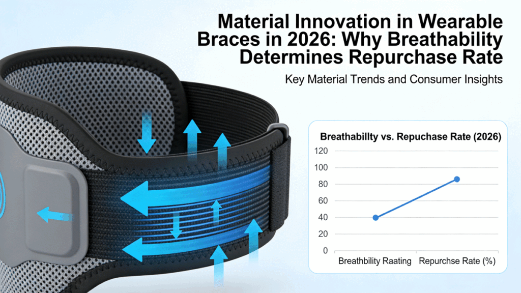
Five years ago, what were the criteria for evaluating a smart knee brace or medical brace? Motor torque, battery life, and correction algorithms. But today, in 2026, as we stand at the intersection of rehabilitation engineering and smart wearables, the key factor determining whether users are willing to repurchase or wear the device long-term after their initial experience has returned to the most basic and fundamental indicator—breathability. This is a very interesting industry turning point: “powerful functionality” is shifting from a “premium” to an “entry barrier,” while “unobtrusive wearing” is the new competitive advantage. The Gap from “Functional Add-ons” to “Skin Extension” For a long time, medical rehabilitation braces and exoskeletons have faced a huge paradox: users often buy them because of essential needs (such as post-operative rehabilitation and sports protection), but abandon them because they are “too uncomfortable.” Traditional braces, in order to achieve support, typically use sealed plastic shells or composite gels, a design that is thermodynamically disastrous. Skin, as the body’s largest heat dissipation organ, becomes trapped in non-breathable materials for an hour, leading to sweat buildup, keratinization, and consequently skin inflammation, odor, and even bacterial infection. For chronically ill patients or elderly users who need to wear them for more than 8 hours a day, this “torture” experience directly results in a large number of devices being left idle. In 2026, this pain point is being completely resolved by breakthroughs in materials science. The key to repurchase rates lies in the micron-scale structure of new materials. Two Major Technological Paths in 2026: Active Temperature Regulation and Ultra-thin Breathability At the beginning of this year, two research findings published in top international journals pointed the way for the industry. 1. The Era of “Active Ventilation” in Smart Textiles A study published in Nature by Professor Tao Xiaoming’s team at Hong Kong Polytechnic University shows us the possibility of brace materials transitioning from “passive breathability” to “active temperature regulation.” The team developed a soft magnetorheological textile with a diameter of only 57 micrometers. It is not only lightweight and flexible, but more importantly, it achieves intelligent adjustment of breathability. This means that future smart braces will no longer be static, dead-cavity structures. By driving the fiber structure to deform through an electrically controlled magnetic field, the brace can sense the skin’s microclimate: when it detects increased temperature or excessive humidity, the microstructure inside the material actively opens, achieving active ventilation. This “breathable” smart material elevates the temperature and humidity comfort of wearable braces to a new level, solving the fatal flaw of traditional braces being “stuffy.” 2. The “Ultra-Thin Revolution” in Interface Materials If magnetorheological fibers solved the breathability problem of macroscopic structures, then the achievements of Professor Wang Yan’s team at the Guangdong Technion-Israel Institute of Technology, published in Science Advances, have solved the “invisibility” problem of the skin-electrode interface. The team developed a 2.7-micrometer-thick ultra-thin hydrogel electrode that is not only resistant to freezing and drying, but also possesses excellent breathability. In wearable braces, the contact surface between the sensor and the skin is often a major area causing inflammation. When the thickness of the interface material is reduced to the sub-micron level, its bending stiffness is significantly reduced, allowing the electrodes to conform to the human body like skin, while allowing sweat to pass through, greatly reducing motion artifacts and skin allergies during long-term monitoring. When users can wear it continuously for 8 days without feeling its presence, repurchase becomes a natural choice. Objective Data: The Economics Behind Breathability Let’s look at some research data from Simon Fraser University in the field of prosthetic sockets: By using 3D-printed lattice structures (similar to honeycomb or trabecular bone), the new sockets improve energy absorption by 1600% when standing compared to solid structures. This porous lattice filling not only brings extreme lightweighting but also creates a huge heat dissipation surface area. Behind this lies an objective fact: breathability is directly related to user compliance. Every 10% increase in compliance leads to an exponential change in the percentage reduction in rehabilitation time, the frequency of consumable replacements, and user trust in the brand. In a market with fierce competition for existing customers, if brace A keeps a user’s skin dry while brace B causes rashes, no matter how advanced B’s algorithm is, users will vote with their feet. In conclusion, as practitioners deeply involved in this field, we must be keenly aware that consumers in 2026 will be sufficiently discerning in the consumer electronics market. They want the support of an exoskeleton, but also the comfort of a pure cotton T-shirt. The industrialization approach of the Hong Kong Polytechnic University team is highly commendable—using commercial-grade raw materials and mature processing techniques to ensure rapid technology deployment. This reminds us that the revolution in breathability cannot remain confined to laboratory samples; it must be integrated into mass production lines at an affordable cost through engineering. The leading players of the future will undoubtedly be those companies that make braces feel like a “second skin” rather than “mechanical armor.” Because only by respecting the skin can we win hearts; only by winning hearts can we win repeat purchases. #Orthopedics #KneeBrace #MedicalDevices #Rehabilitation #HealthcareInnovation #MedicalDistributor

Scroll to Top