SAK BRACE

SAK BRACE specializes in medical orthopedic braces, rehabilitation supports, and customized OEM solutions for global distributors, clinics, and healthcare brands.

WhatsApp: 0086-13343185033 / 0086-13132404899 Email: info@sakbrace.com

Author name: admin

Uncategorized

ACL Reconstruction Rehabilitation Timeline: Stages, Milestones, and Return to Sport

Rehabilitation after anterior cruciate ligament (ACL) reconstruction is a lengthy and structured process, typically lasting 9 to 12 months. The core goal of rehabilitation is not solely based on time, but rather on a criterion-based progression strategy, meaning that the next stage can only proceed after specific functional indicators are achieved. This article systematically describes the rehabilitation timeline and key points of each stage after ACL reconstruction based on current evidence-based medicine. Preoperative Stage Increasing evidence suggests that preoperative rehabilitation has a positive impact on postoperative outcomes. Accelerated recovery programs typically incorporate the preoperative stage into the overall rehabilitation framework. The focus of preoperative rehabilitation is on controlling joint inflammation, restoring knee joint range of motion, activating quadriceps function, and preparing patients psychologically and academically for postoperative rehabilitation. Studies show that patients who begin systematic rehabilitation training preoperatively experience faster early postoperative functional recovery. Postoperative Weeks 0-2: Acute Protection Phase This stage marks the beginning of rehabilitation, with core tasks revolving around wound healing, inflammation control, and the restoration of basic function. Immediately after surgery, the following aspects need attention: First, control pain and swelling through ice application, limb elevation, and ankle pumps; second, restore full knee extension as early as possible with brace support, which is crucial to prevent postoperative flexion contracture; third, activate the quadriceps to prevent early muscle atrophy. Regarding weight-bearing, simple ACL reconstruction usually allows for tolerable weight-bearing; if meniscus repair is combined, a 6-week restriction of no weight-bearing or limited weight-bearing is required. Patients typically need to use crutches for 2 to 3 weeks during this stage. Weeks 2-6 post-surgery: Early Range of Motion Recovery During this stage, the focus of rehabilitation shifts from simple protection to the systematic recovery of range of motion. Knee flexion angle should typically gradually reach over 90°. Patients should gradually transition to a normal gait pattern during this stage, progressing from partial weight-bearing to full weight-bearing walking. Quadriceps strengthening training needs to be continuously promoted, with the goal of achieving over 60% of the strength of the unaffected quadriceps by the end of this stage (measured by the limb symmetry index). Neuromuscular electrical stimulation can be used as an adjunct for early muscle strength recovery. Activities of daily living gradually resume during this stage. Office-based sedentary work can usually be resumed 4 to 6 weeks post-surgery; driving should be considered based on specific advice and insurance terms, generally 4 to 6 weeks post-surgery. Weeks 7-9 Post-Surgery: Mid-Term Intensive Training Prerequisites for entering this stage include: knee range of motion reaching 0° extension and 115° flexion or more, joint effusion controlled below 1+, and gait returning to normal. Training focus shifts to balance training, neuromuscular re-education, and aerobic exercise. For strength training, the goal is to increase the quadriceps strength symmetry index to over 70%. Closed-chain and open-chain exercises should be introduced gradually under the guidance of a physician to avoid excessive stress on the reconstructed graft. Weeks 10-16 Post-Surgery: Late-Term Functional Training This stage is marked by the introduction of running. The prerequisite for starting running is a quadriceps strength symmetry index of over 80%. The training content transitions from simple strength training to a more intensive gym-based basic training program, while emphasizing standardized training in landing mechanics and movement patterns. The target range for the strength symmetry index at this stage is 75% to 80%. 4-6 Months Post-Surgery: Transition Period This is a crucial stage in rehabilitation, transitioning from basic function to motor function. Training content introduces jumping, sprinting, deceleration, and change-of-direction agility training. The core goal at this stage is to achieve a quadriceps strength symmetry index and a single-leg jump test symmetry index of over 85%. Research indicates that 3-6 months post-surgery is a critical window for exercise load programming. 6-12 Months Post-Surgery: Return to Exercise Phase The final stage of rehabilitation focuses on sports-specific training and physical preparation. Medical clearance for returning to competition requires the following conditions: no joint pain or effusion; quadriceps strength symmetry index and jump test symmetry index both exceeding 90%; and an assessment of the patient’s confidence and psychological readiness for exercise. It is important to note that even after clearance for return, continuous strength and neuromuscular training is necessary for 12 months post-surgery to minimize the risk of re-injury. Graft Type and Rehabilitation Differences Different graft types have a certain impact on the rehabilitation process. Bone-patellar tendon-bone grafts, hamstring tendon grafts, quadriceps tendon grafts, and allogeneic grafts differ in the load intensity and progress at each stage of rehabilitation. In clinical practice, rehabilitation plans need to be individualized according to the specific graft type and surgical procedure. Conclusion ACL reconstruction rehabilitation is a standard-guided, phased process, typically lasting 9 to 12 months. The timeframes for each stage are only a reference framework; the actual progress needs to be dynamically adjusted based on the individual patient’s functional recovery. Under the joint guidance of professional physical therapists and surgeons, and using a structured, progressive rehabilitation plan, the vast majority of patients can safely return to their pre-injury motor skill levels.

Uncategorized

ACL Injury Grades Explained: Grade 1, Grade 2 & Grade 3 ACL Tears

Introduction The anterior cruciate ligament (ACL) is one of the key stabilizing structures within the knee joint and plays an essential role in controlling anterior tibial translation and rotational stability. ACL injuries are among the most common knee injuries, particularly in sports involving sudden stops, pivoting, jumping, and rapid changes of direction. However, not all ACL injuries are the same. The severity can range from a mild ligament sprain with minimal structural damage to a complete rupture causing significant knee instability. Understanding ACL injury grades helps clinicians evaluate injury severity, define rehabilitation goals, and determine whether additional support, including functional bracing, may be beneficial during recovery. Clinically, ACL injuries are commonly classified into three grades: ACL Injury Grade Injury Description Ligament Condition Knee Stability Grade 1 Mild sprain Ligament fibers stretched with minimal microscopic damage Stable Grade 2 Partial tear Partial disruption of ACL fibers Mild to moderate instability Grade 3 Complete rupture Full ligament tear with loss of functional stability Significant instability This article explains the differences between Grade 1, Grade 2, and Grade 3 ACL injuries, including clinical symptoms, treatment considerations, and the potential role of functional ACL braces during rehabilitation. 1. What Are ACL Injury Grades? ACL injury grading is a clinical classification system based on the extent of ligament damage and the resulting effect on knee stability. The grading process considers several factors, including: Common clinical tests include: MRI is frequently used as an additional diagnostic tool to assess ACL integrity and identify associated injuries such as: Although imaging provides important structural information, treatment decisions are not based solely on MRI findings. Functional stability, patient goals, and activity demands are equally important. 2. Grade 1 ACL Injury: Mild Sprain Definition A Grade 1 ACL injury represents the mildest form of ACL damage. The ligament is overstretched but remains structurally intact. Only a small number of microscopic fibers may be affected, and the ACL can still provide normal mechanical stability to the knee. Unlike higher-grade injuries, Grade 1 ACL sprains usually do not result in significant ligament laxity. Typical Symptoms Patients with Grade 1 ACL injuries may experience: Symptom Clinical Presentation Pain Mild discomfort around the knee Swelling Usually limited or temporary Stability Knee remains functionally stable Daily activities Walking is generally unaffected Sports activity Discomfort may appear during running, jumping, or sudden movements Most patients do not experience a feeling of the knee “giving way.” Treatment Approach Conservative management is typically recommended for Grade 1 ACL injuries. Early treatment may include the PRICE principle: As symptoms improve, rehabilitation focuses on: Recovery is generally faster compared with higher-grade injuries, and many patients return to daily activities within several weeks. Role of ACL Bracing For most Grade 1 ACL injuries, functional ACL braces are not routinely required because the ligament maintains adequate stability. However, some patients may choose to use a lightweight knee support during the later stage of rehabilitation or return-to-sport phase to: The decision should always be based on individual patient needs rather than injury grade alone. 3. Grade 2 ACL Injury: Partial ACL Tear Definition A Grade 2 ACL injury involves partial disruption of the ligament fibers. Some ACL fibers remain intact, but the ligament becomes elongated and loses part of its normal tension. Compared with Grade 1 injuries, Grade 2 tears are more likely to affect knee stability, especially during rotational movements or high-demand activities. Typical Symptoms Symptom Clinical Presentation Pain Moderate pain, especially during movement Swelling More noticeable swelling or joint effusion Stability Mild to moderate instability Movement Difficulty with pivoting, cutting, or sudden direction changes Functional symptoms Possible “giving way” episodes Patients often report instability during: Treatment Approach Management of Grade 2 ACL injuries requires individualized assessment. Conservative Rehabilitation For patients with acceptable knee stability, rehabilitation may focus on: A structured rehabilitation program may take approximately 3–4 months, depending on patient progress. When Surgery May Be Considered Surgical intervention may be discussed when patients have: Treatment decisions should consider: Role of Functional ACL Bracing Grade 2 ACL injuries represent one of the areas where functional bracing is frequently considered. A properly designed ACL functional brace may help by: However, brace selection should be based on: A brace should complement rehabilitation rather than replace strength and neuromuscular training. 4. Grade 3 ACL Injury: Complete ACL Rupture Definition A Grade 3 ACL injury represents a complete rupture of the anterior cruciate ligament. The ligament fibers are fully disrupted, resulting in significant loss of mechanical stability. The rupture may occur: Typical Symptoms Symptom Clinical Presentation Injury sensation Possible “pop” feeling or sound at injury moment Swelling Rapid and significant swelling due to hemarthrosis Stability Marked instability and knee giving way Activity limitation Difficulty with pivoting and high-demand sports Motion Possible limitation due to pain and swelling Many patients experience difficulty trusting the injured knee during daily movement. Treatment Approach Treatment for Grade 3 ACL injuries is highly individualized. Non-Surgical Management Conservative treatment may be appropriate for: A structured rehabilitation program focuses on: ACL Reconstruction Surgery ACL reconstruction may be considered for patients who: The goal of ACL reconstruction is to restore knee stability and reduce the risk of secondary damage, including: Importantly, not every Grade 3 ACL tear automatically requires surgery. Modern ACL management emphasizes patient-specific decision-making rather than injury grade alone. 5. ACL Injury Grades Comparison: Grade 1 vs Grade 2 vs Grade 3 Although ACL injuries are commonly classified into three grades, the actual clinical situation is more complex than simply matching injury grade with treatment choice. Each grade represents a different level of structural damage and functional impact. The following table provides a general comparison: Comparison Factor Grade 1 ACL Injury Grade 2 ACL Injury Grade 3 ACL Injury Ligament Damage Mild stretching or microscopic fiber damage Partial tearing of ACL fibers Complete rupture of ACL Structural Integrity Mostly preserved Partially compromised Completely disrupted Knee Stability Stable Mild to moderate instability Significant instability Swelling Usually mild Moderate swelling possible Often rapid and significant swelling Functional Symptoms Mild discomfort during activity Possible

Uncategorized

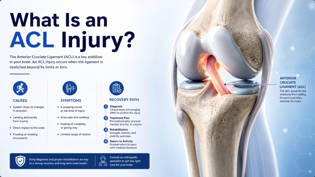
What is an Anterior Cruciate Ligament (ACL) Injury?

I. Understanding the Anterior Cruciate Ligament (ACL)The anterior cruciate ligament (ACL) is one of the four main ligaments in the knee joint, located in the center of the knee. It originates from the posterior aspect of the lateral intercondylar fossa of the femur and attaches anteriorly, distally, and medially to the anterior part of the intercondylar spine of the tibial plateau. Together with the posterior cruciate ligament (PCL), it forms the cruciate ligaments of the knee, crossing in an “X” shape at the front of the knee joint. The core function of the ACL is to prevent excessive anterior displacement of the tibia relative to the femur, while also providing rotational stability to the knee joint. In short, the ACL acts as a “safety lock” for maintaining the dynamic stability of the knee joint—it constantly restricts the range of motion of the knee joint during running, jumping, sudden stops, and changes of direction, preventing abnormal joint displacement. II. Epidemiology: A Number Not to Be IgnoredACL injuries are among the most common orthopedic sports injuries. According to a long-term population-based study in the United States, the annual incidence of ACL injuries is approximately 68.6 per 100,000 person-years. In the United States, approximately 250,000 ACL injuries occur annually, with over 175,000 requiring ACL reconstruction surgery. Other literature estimates between 100,000 and 200,000 ACL tears annually in the US. The incidence of ACL injury in the general population is approximately 1 in 3,500, equivalent to about 95,000 new ACL ruptures each year. In terms of age distribution, athletes aged 15 to 25 account for over 50% of ACL injuries. A survey of active athletes in my country found an overall incidence of ACL rupture of 0.43%; among professional female athletes, the incidence was 0.71%, and for males, it was 0.29%, with women approximately 2.37 times more likely to be affected than men. The Institute of Sports Medicine at Peking University Third Hospital currently performs over 1,400 ACL reconstruction surgeries annually. III. Why do ACL injuries occur? Over 70% of ACL injuries are caused by non-contact forces. Typical scenarios include: sudden deceleration and change of direction (crossover), turning while the foot is fixed, improper landing posture, and sudden stops. Domestic data shows that 78% of ACL injuries are non-contact injuries. Specifically, common mechanisms of ACL injury include: Sudden stop and change of direction: Sudden deceleration and change of direction during movement. Single-leg landing: Landing with the entire foot to stabilize the tibia, resulting in valgus displacement when the knee is in a small flexion position. Knee hyperextension: Excessive extension of the knee joint, causing excessive stress on the ACL. Direct impact: Direct force to the knee joint from the side (such as a tackle in soccer). ACL injuries are rare. Approximately 50% of ACL injury patients also have meniscus tears; more than two-thirds of complete ACL ruptures are accompanied by meniscus and/or articular cartilage damage. In acute injuries, the lateral meniscus is more commonly involved; in chronic ACL injuries, medial meniscus injuries are more common. IV. Symptoms and Diagnosis When an ACL injury occurs, patients often hear or feel a “pop” sound within the knee joint. Subsequent symptoms include: Severe pain, inability to continue activity Rapid swelling of the knee joint (usually occurring within minutes to 3 hours after injury, primarily due to effusion) Limited range of motion A feeling of “giving way” or instability in the knee when bearing weight It is noteworthy that 70% of acute knee injuries with effusion also involve ACL injury. For diagnosis, the Lachman test is the most effective method for examining ACL injuries. MRI has an accuracy rate of up to 95%, clearly showing ligament damage and simultaneously assessing accompanying meniscus and cartilage damage. V. Treatment: Conservative or Surgical? Treatment plans for ACL injuries need to be individualized based on the patient’s age, activity level, and injury severity. Conservative treatment is suitable for patients with milder injuries (Grade I) or lower daily activity requirements (e.g., International Documentation Committee on Knee Classification III-IV). Treatment includes protection, rest, ice application, compression bandages, elevation of the affected limb (RICE principle), and systemic physical therapy. Surgical treatment—ACL reconstruction—is the preferred option for patients with complete ACL rupture or those wishing to return to high-intensity sports. The surgery typically employs arthroscopic techniques, using tendons from other parts of the patient’s body (autologous hamstring tendons or bone-patellar tendon-bone) or allogeneic tendons to replace the damaged ligament. The timing of surgery is generally recommended within 3 months of injury to reduce the risk of secondary cartilage and meniscus damage. In terms of long-term outcomes, the success rate of ACL reconstruction is between 75% and 95%. However, less than 50% of patients can return to normal athletic performance within one year post-surgery. For professional soccer players, over 92% can return to the field after surgery, and approximately 80% can recover to their pre-injury level. VI. Long-term effects: More than just a ligament problem The consequences of ACL injury extend far beyond the ligament itself. Regardless of whether surgery is performed, patients with ACL injuries have a significantly increased risk of developing knee osteoarthritis. Approximately 50% of patients with ACL injuries will develop varying degrees of osteoarthritis within 10 to 20 years of the initial injury. In the long term, the odds ratio for total knee replacement due to osteoarthritis in patients with ACL injuries has increased nearly sevenfold. VII. Prevention: The Best TreatmentNeuromuscular training has been proven to effectively reduce the risk of ACL injuries, especially for female athletes. Training includes strengthening core muscles (hips, pelvis, lower abdomen), improving biomechanical patterns during lower limb landing, and strengthening hamstrings. Studies have shown that neuromuscular training several times a week for more than six weeks can significantly reduce non-contact ACL injuries in female athletes. It is important to note that preventative knee braces do not prevent ACL injuries—true protection comes from scientific training and correct movement patterns. Anterior cruciate ligament (ACL) injury is one of the most concerning injuries in sports medicine. It commonly affects young,

Uncategorized

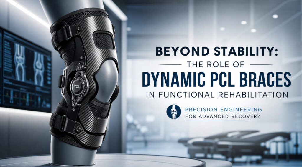
Beyond Stability: The Role of Dynamic PCL Braces in Functional Rehabilitation

In traditional orthopedic rehabilitation, knee braces are often seen as auxiliary devices for protection and stability—their core functions are to restrict movement, protect healing tissue, and provide mechanical stability. However, in the modern orthopedic rehabilitation perspective, the role of braces has far surpassed simple fixation and immobilization. The goal of rehabilitation is no longer “immobility,” but “how to move better.” This shift in concept not only redefines the value of braces but also fundamentally changes the rehabilitation pathway for posterior cruciate ligament (PCL) injuries. Part One: From Immobilization to Controlled MovementIn the past, PCL injury rehabilitation relied on static braces or plaster casts. The core logic of these braces is simple and direct: restrict all unnecessary movement, protect damaged or reconstructed ligament tissue, and prevent posterior tibial displacement—the most fundamental biomechanical problem of the PCL. However, excessive restriction of movement itself brings a series of new problems. Studies have shown that prolonged immobilization for more than 6 weeks can lead to a joint stiffness rate as high as 68% and quadriceps atrophy of more than 40%. Muscle atrophy, decreased joint mobility, and delayed functional recovery—these complications run counter to the ultimate goal of rehabilitation. It is against this backdrop that modern rehabilitation concepts have shifted from “absolute immobilization” to “controlled movement.” Rehabilitation is no longer about restricting all movement, but about restoring function within safe limits—allowing patients to move under protection and rehabilitate through movement. This shift means that the role of braces has evolved from “preventing movement” to “guiding movement.” Part Two: The Value of Dynamic Support in PCL RehabilitationThe core biomechanical issue of the PCL is posterior tibial translation. The primary function of the PCL is to limit posterior displacement of the tibia relative to the femur, a role particularly crucial in knee flexion. Therefore, the core task of PCL rehabilitation braces is not simply “fixing the knee joint,” but effectively controlling posterior tibial translation. The value of dynamic braces lies precisely here. Unlike the static restrictions of traditional rigid braces, dynamic braces actively counteract the tendency for posterior tibial translation by applying a controllable, forward-oriented force to the lower leg during knee flexion. Biomechanical studies have confirmed that dynamic bracing can reduce posterior tibial displacement under stress from an average of 7.1 mm to 2.68 mm (p<0.001). In a PCL disarticulation specimen model, a dynamic brace applying a 50 N anterior force also significantly reduced posterior tibial displacement (P=0.032). The true significance of dynamic bracing lies in the fact that it does not simply “lock” the knee joint, but provides continuous biomechanical support while allowing joint movement. This allows patients to begin functional activities early in rehabilitation—from standing and sitting to climbing stairs and squatting—without worrying about the risk of secondary injury from posterior tibial displacement. Part Three: The Balance Between Protection and FunctionRehabilitation always faces a core contradiction: the tension between overprotection and premature mobilization. Overprotection means adequate tissue healing, but at the cost of muscle atrophy, joint stiffness, and delayed functional recovery. Excessive mobilization may improve joint range of motion and muscle strength recovery, but may increase the stress burden on the graft or healing tissue. The challenge of PCL rehabilitation lies precisely in finding the optimal solution to this contradiction. Dynamic braces offer a balanced solution. On one hand, they effectively protect the PCL from excessive stress caused by posterior tibial displacement through continuous anterior support; on the other hand, they allow for safe flexion and extension of the knee joint, supporting 0-90° of joint range of motion in the early postoperative period. Studies show that rehabilitation programs assisted by dynamic braces can restore 85%-92% of joint range of motion after 6 weeks, and quadriceps muscle strength increases 3.2 times compared to the acute phase. Protection aims to better restore function, and functional recovery cannot be separated from appropriate protection—dynamic braces bridge this gap. Part Four: Patient-Centered PCL Brace Selection Modern orthopedic rehabilitation emphasizes “patient-centered care”—meaning that brace selection cannot be a one-size-fits-all approach. PCL brace selection requires comprehensive consideration of multiple factors: the severity of the injury (simple PCL injury or multi-ligament injury), whether surgical treatment is required and the surgical method, the current stage of rehabilitation (acute phase, rehabilitation phase, or functional recovery phase), the patient’s daily activity level and exercise needs, and the patient’s adherence to the brace. Different patients have different brace needs at different stages—stronger tibial support is needed in the acute phase, greater freedom of movement is required during rehabilitation, and a new balance needs to be struck between protection and training during functional recovery. The era of “One brace fits all” is over. A brace solution precisely tailored to the individual needs of each patient is key to achieving optimal rehabilitation outcomes. In conclusion, PCL braces are no longer merely tools to restrict unnecessary movement. Their core value lies in providing controlled support, helping patients safely navigate every stage of rehabilitation—from postoperative immobilization to early mobilization, from basic training to functional reconstruction. The future of orthopedic braces lies in the organic integration of biomechanical principles, clinical understanding, and patient needs. At SAK BRACE, we continue to focus on developing orthopedic solutions that align with this philosophy, supporting clinicians’ professional decision-making and improving patient rehabilitation outcomes.

Uncategorized

The Two Ends of the “Balance” in Postoperative Rehabilitation: Clinical Selection of Static vs. Dynamic PCL Braces

In the field of orthopedic sports medicine, the management of posterior cruciate ligament (PCL) injuries has always been a challenging issue. Compared to the anterior cruciate ligament (ACL), the PCL has a more robust anatomy, a more complex blood supply, and its injuries are often accompanied by damage to the complex structures of the posterolateral or posteromedial horns. During surgical treatment or conservative rehabilitation, the use of braces is considered one of the core variables determining the quality of ligament healing and the recovery of joint function. Currently, a key choice in clinical practice is: during the patient’s long rehabilitation period, should the traditional static brace be chosen, or the dynamically brace that has gained popularity in recent years? To objectively examine this issue, it is first necessary to clarify the design philosophy and mechanical mechanism of both. A static brace, as the name suggests, primarily functions to provide “rigid restraint.” These types of braces typically use a rigid polymer frame, combined with hinges and padding, to physically restrict posterior displacement of the tibia relative to the femur. Their greatest advantage lies in providing definitive protection. In the early postoperative period (0-6 weeks), when the graft or suture site is still in its weakest mechanical window, static braces, by locking the extension angle or applying anterior corrective forces, can effectively prevent tibial subsidence caused by gravity, thus creating a stable tension environment for the collagen fiber bridging of the PCL. However, static braces also have significant limitations. Prolonged rigid fixation can easily lead to arthropathic muscle inhibition (AMI), resulting in significant atrophy and decreased strength of the quadriceps during immobilization. Since the active contraction of the quadriceps is the core power source for maintaining tibial position after PCL injury, static braces, while protecting the ligaments, objectively “blunt” proprioceptive input, delaying the recovery process of neuromuscular control. In contrast, dynamic braces represent an evolution in rehabilitation philosophy. Dynamic braces are not simply “bindings,” but rather provide variable corrective forces that change with the angle during knee flexion and extension through elastic elements or adjustable air pressure systems. Their core concept is “protection during movement.” For example, some dynamic braces use elastic bands placed on the back of the lower leg to generate a gradually increasing forward pull as the patient actively flexes the knee, mimicking the synergistic contraction of the hamstrings. This design allows patients to perform open-chain or closed-chain movements earlier, significantly reducing the inhibitory effect on the quadriceps and effectively avoiding the vicious cycle of “immobilization-atrophy-re-injury.” However, do dynamic braces have an absolute advantage? Based on current evidence-based medicine, the answer is not unanimous. Dynamic braces require a high level of patient understanding and compliance. If the patient cannot accurately understand the logic of using the brace, or if their gait is uncoordinated during walking, the variable forces provided by the dynamic brace may actually become an unstable source of interference. Furthermore, dynamic braces are typically much more expensive than standard static braces, and their corrective effect is highly dependent on the precise adjustments made by the rehabilitation therapist, which limits their widespread adoption in primary healthcare institutions. From a clinical decision-making perspective, the key variables in choosing a brace are the severity of the injury and the stage of rehabilitation. In the acute postoperative period (0-4 weeks), when there is significant joint effusion and inflammation, the “absolute stillness” provided by a static brace is an irreplaceable safety guarantee. However, in the mid-to-late stages of rehabilitation (after 8 weeks), when ligaments have begun to connect with scar tissue, introducing a dynamic brace, combined with proprioceptive training, can effectively promote fibrous alignment during graft ligamentization and reduce the risk of articular cartilage degeneration due to prolonged immobilization. Objectively speaking, static and dynamic braces are not “replacements” but rather complementary. Modern orthopedic rehabilitation tends to adopt a “phased switching” strategy: immediately after vital signs stabilize, use an adjustable-angle static brace for early protection; once neuromuscular control has recovered to a critical level, transition to a dynamic brace to enhance functional recovery. Clinicians should not blindly pursue novel technologies, but rather develop individualized brace prescriptions based on patients’ body mass index (BMI), bone mineral density, occupational needs, and psychological expectations. Regardless of technological advancements, braces are ultimately just assistive tools. The core of PCL rehabilitation lies in high-quality physical therapy intervention. Only under the guidance of professional muscle strength assessment and gait analysis can the stability of static braces and the flexibility of dynamic braces complement each other, ultimately helping patients achieve the essential leap from “structural healing” to “functional healing.” Orthopedics #PCLBrace #SportsMedicine #OrthopedicRehabilitation #MedicalDevices

Uncategorized

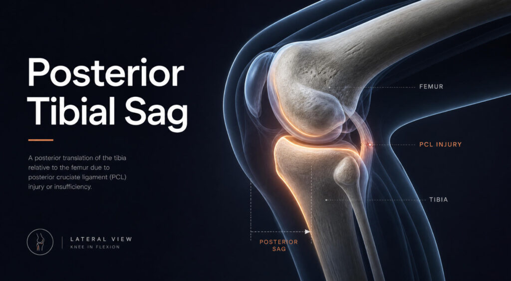
Posterior Tibial Sag Sign: The Most Reliable Clinical Clue for PCL Injury

The posterior tibial sag sign is one of the most typical clinical signs of posterior cruciate ligament (PCL) injury in the knee joint. It visually reflects the abnormal posterior displacement of the tibia relative to the femur under gravity after ligament damage. Understanding the “Stabilizing Force of the Knee”: The Posterior Cruciate Ligament To understand posterior tibial sag, it’s essential to first understand the posterior cruciate ligament (PCL). The PCL is the strongest ligament in the knee joint, approximately twice the strength of the anterior cruciate ligament (ACL). Its core function is to prevent posterior dislocation of the tibia relative to the femur, providing about 95% of the resistance against the force of posterior tibial displacement. You can think of it as a strong rein, firmly “tying” the tibia of the lower leg to the femur of the thigh, ensuring knee stability during flexion and extension. How does the sag sign occur? When this powerful “reins”—the PCL—ruptures, the tibia loses its primary constraint on posterior movement. The posterior cruciate ligament (PCL) injury is most commonly seen in a scenario known as a “dashboard injury.” For example, in a traffic accident, when the knee is bent, the lower leg violently impacts the car’s dashboard, and this backward force forces the tibia to shift backward, leading to a tear in the PCL. After the ligament tears, if the patient lies flat with their knee bent at 90 degrees, the unrestrained upper tibia will naturally “sink” or “drop” backward due to gravity. This is the root cause of “posterior tibial subsidence.” How to detect this sign? The examination method for this sign is relatively simple and is a commonly used physical examination technique by orthopedic and sports medicine doctors. Examination position: The patient lies supine with both hip and knee joints bent at 90 degrees and both feet flat on the examination table. Observation and comparison: The examiner observes the contour of the patient’s knee joint from the side. Normally, due to the traction of the posterior cruciate ligament (PCL), the tibial plateau (the plane at the upper end of the tibia) should be located about 1 cm anterior to the femoral condyle (the lower end of the femur), forming a small “step.” Positive result: If the PCL is ruptured, this “step” disappears, and the tibial plateau shifts significantly posteriorly, becoming level with or even posterior to the anterior edge of the femoral condyle. This sinking phenomenon is more pronounced when compared to the healthy side (the uninjured leg). Clinical significance and accuracy of the “sinking sign”: The “posterior tibial sinking sign” is a highly specific indicator for diagnosing PCL injuries. Studies show that the diagnostic specificity of this sign can reach 100%, meaning that if this sign is positive, a PCL injury is almost certain. Its diagnostic sensitivity is approximately 79%, indicating that most patients will present with this sign, but a small number may have atypical symptoms. Clinically, doctors often use this test in conjunction with the posterior drawer test. The latter involves the examiner actively pushing the tibia backward to assess the degree of posterior displacement. Based on the distance of posterior tibial displacement, the injury can be graded: Grade I: Displacement less than 5 mm. Grade II: Displacement between 5 and 10 mm. Grade III: Displacement greater than 10 mm. What to do if “sinking” is found? If “posterior tibial sinking” is found during a physical examination, the doctor will usually recommend an MRI. MRI can clearly show whether the posterior cruciate ligament is partially torn or completely ruptured, and simultaneously assess whether other structures within the knee joint (such as the meniscus and cartilage) are damaged. The treatment plan depends on the severity of the injury. For simple posterior cruciate ligament tears without other accompanying injuries, conservative treatment is usually the first choice. The core of the treatment plan is strengthening the quadriceps (the muscles on the front of the thigh). Strong quadriceps can compensate to some extent for the stabilizing function of the damaged ligament, helping patients restore functional stability to the knee joint. In summary, the “posterior tibial subsidence sign” is an important indicator of posterior knee joint stability. It is simple and intuitive, serving as a key basis for doctors to determine whether the posterior cruciate ligament is damaged. Understanding this sign helps us comprehend the mechanisms of knee injuries and recognize the importance of timely and professional diagnosis for subsequent treatment and rehabilitation.

Category 1

From Rehabilitation to Prevention: An Objective Examination of the Value of Braces in Sports Injuries

In today’s nationwide fitness boom, sports injuries have become an almost inevitable consequence of this enthusiasm, whether for marathon runners, weightlifting enthusiasts, or amateur athletes. As a practitioner/enthusiast focusing on sports rehabilitation, I’ve recently found in discussions with colleagues that opinions on braces are often polarized: some see them as a lifeline after injury, while others consider them a restrictive burden. Today, I want to discuss the topic of “Common Sports Injuries and Braces” objectively, without subjective bias. I. The Core Role of Braces: Not Healing, But Managing First, we need to clarify a concept: braces themselves do not possess healing capabilities; they cannot heal torn ligaments or eliminate inflammation. Their core function is biomechanical intervention and management. At different stages of sports injuries, braces play three distinct roles: 1. Immobilization Phase: After the acute phase of the injury or post-operatively, braces restrict movement in specific directions, providing a tension-free and stable environment for soft tissue healing. 2. Functional Phase: As tissues begin to heal, a balance needs to be struck between protection and mobility. Functional braces allow joints to move within a safe range, preventing muscle atrophy and joint stiffness. 3. Prophylactic Phase: This is the most controversial yet most widely used area, where braces are used by healthy individuals or those with prior injuries to prevent injury or recurrence during high-risk sports. II. Objective Analysis of Common Injuries and Corresponding Braces Let’s look at some common examples in sports scenarios: 1. Knee Joint: Anterior Cruciate Ligament (ACL) Injury Common Scenarios: Sudden stops and changes of direction in basketball and soccer. Brace Application: Hinged knee braces are standard in post-operative rehabilitation. It controls the extension angle, preventing overextension that strains reconstructed ligaments. Objective Perspective: For ACL injuries, the academic community generally agrees that postoperative bracing is part of the rehabilitation process. However, the evidence for the effectiveness of prophylactic braces in preventing first-time injuries is insufficient. It may give the wearer a false sense of invulnerability, leading to more dangerous movements. 2. Ankle: Recurrent Sprains Common Scenarios: Running, badminton, outdoor hiking. Brace Application: Semi-rigid or soft ankle braces. Objective Perspective: Compared to the knee, ankle braces have more robust evidence for prevention. For individuals with a history of multiple sprains, wearing a brace during high-intensity activities (such as contact sports and trail running) can significantly reduce the risk of re-sprains. However, it should not replace proprioceptive training—the brain’s control over ankle position is the ultimate “ankle protection.” 3. Wrist: Scaphoid Fracture / Triangular Fibrocartilage Complex Injury Common Scenarios: Falling and landing on the palm (skiing, skateboarding), gymnastics. Brace Application: Wrist immobilization brace. Objective Perspective: Wrist braces are often synonymous with “absolute immobilization.” Due to the unique blood supply to the scaphoid bone, healing is slow, requiring high compliance with braces. Many enthusiasts remove the brace themselves once the pain subsides, often leading to nonunion—a very serious misconception. 4. Lower Back: Spondylolysis / Lumbar Disc Herniation Common Scenarios: Weightlifting, golf, sudden physical activity after prolonged sitting at a desk. Brace Application: Rigid or soft lumbar support. Objective Perspective: The core function of a lumbar brace is to increase intra-abdominal pressure and distribute the load on the lumbar spine. It is an excellent aid during extreme weight-bearing activities (such as heavy squats). However, prolonged reliance on lumbar support in daily life can lead to core muscle atrophy and weakening, making the lumbar spine more vulnerable. III. Three Objective Suggestions for Sports Enthusiasts Based on the above observations, I have three preliminary thoughts for those considering or currently using braces: 1. Understand the “Expiration Date” of Braces Braces are “crutches” in the rehabilitation process, not permanent “prosthetics.” Except for a few athletes with special needs, most people’s ultimate goal should be to get rid of braces and regain joint control through rehabilitation training. 2. Beware of “Brace Dependence” and “Brace Illusion” On the one hand, over-reliance on braces can weaken proprioception; on the other hand, the sense of security provided by wearing braces may induce actions beyond the current joint’s capacity, causing more insidious injuries. 3. The Importance of Personalized Customization There is a wide variety of sports braces on the market. For simple sprains, a general-purpose brace may suffice; however, for complex ligament injuries or post-operative rehabilitation, a brace custom-made by a rehabilitation physician or orthotist provides more precise pressure distribution and range of motion control, far exceeding the value of a regular “protective gear.” In Conclusion Sports injury rehabilitation is an art of “balance.” A brace is a precise ruler in this artistic creation, but it cannot draw the blueprint for health. True freedom of movement comes from strong muscle strength, sensitive neural control, and a scientific training plan. A brace is merely a reliable and rational partner on our journey back to the track. Interactive Topic: Have you ever been injured while exercising? What was your experience with the brace or protective gear you used? Feel free to share your experiences and opinions in the comments section, and let’s exchange tips on avoiding common pitfalls. #SportsRehabilitation #SportsInjury #HealthEducation #PhysicalTherapy #WorkplaceHealth

Category 1

Knee Brace Procurement in 2026: 5 Key Factors Buyers Should Not Ignore

With the continued expansion of the global orthopedic rehabilitation market in 2026, knee braces are no longer simply a combination of “plastic + Velcro.” According to the latest data from Research and Markets, the global knee brace market is projected to reach $2.74 billion in 2026 and is iterating towards digitalization and precision. However, against the backdrop of technological upgrades and increased entrants, many medical institution procurement personnel and rehabilitation physicians are facing a new “choice dilemma.” Should they pursue high-end imported “cutting-edge technology,” or choose more cost-effective domestic emerging products? Should they blindly stockpile to cope with centralized procurement, or refine their approach to match clinical needs? Based on recent bidding and procurement regulations and industry technology trends, this “2026 Knee Brace Procurement Pitfall Avoidance Guide” will help you make more professional business decisions. Pitfall 1: Blindly Believing in “Universal Braces,” Ignoring Precise Clinical Subtyping Avoidance Strategy: From “General-Purpose” to “Indication Segmentation” The most common misconception in procurement is treating knee braces as a single category. In fact, the market segmentation in 2026 is already extremely clear: unloader knee braces for anterior cruciate ligament injuries, osteoarthritis (OA), post-meniscus repair braces, and patellar stabilization braces differ significantly in their biomechanical design. Professional advice: When purchasing products, it is essential to configure them according to the types of surgeries performed and the structure of outpatient cases. For example, for osteoarthritis patients, unloader knee braces can change the force line through a three-point biomechanical design, which is already the mainstream conservative treatment approach internationally. If your purchasing list only includes ordinary hinged braces and ignores these types of braces with “therapeutic functions,” you may miss the growth point in the conservative treatment market. Pitfall Two: Comparing only prices and ignoring “wearability” design Avoidance strategy: Focus on ergonomics and material innovation Materials science in 2026 has revolutionized knee braces. The bulky, hot, and poorly adhered “iron frames” of the past are being replaced by lightweight polymer composite materials and even smart fabrics. If procurement personnel only focus on the few yuan price difference during bidding, ignoring the comfort of wearing the brace, it will ultimately lead to patients being unwilling to wear it and compromising rehabilitation effects, which is undoubtedly the biggest waste. Professional advice: When inspecting goods, not only should you check the qualifications, but also pay attention to the breathability of the lining, the edge treatment of the brace, and the anti-slip design of the straps. Especially for women, the elderly, and athletes, is there sufficient adjustment range? A report from Huanyang Market indicates that adjustable pressure-distributing braces and skin-friendly materials have a much higher repurchase rate than traditional rigid plastic braces. Pitfall Three: Qualification review becomes a formality, neglecting “full-process authorization” Avoidance strategy: Strictly adhere to compliance red lines and rigorously investigate the authorization chain Recent consumable selection announcements issued by hospitals in many regions have repeatedly emphasized a key phrase: “full-process authorization cannot be interrupted.” With the increasingly stringent national supervision of medical devices, especially for high-value consumables and polymer material braces, product registration certificates, production licenses, and authorization at each level of agency must be clearly verifiable. Professional advice: Procurement audits in 2026 will only become more stringent. Before selecting a supplier, it is crucial to verify the following: Product Registration Certificate: Ensure the scope of application aligns with clinical needs. Authorization Chain Integrity: Authorization documents at each level from the manufacturer to the bidding company must be continuous, eliminating “affiliation” and “smuggling.” UDI Traceability Code: Ensure each brace has a unique identifier, enabling full traceability from production to use. Pitfall Four: Neglecting “Digitalization” and “Smart Ecosystem” Integration Capabilities Avoidance Strategy: Focus on the future and consider the equipment’s interface capabilities. According to LP Information’s latest insights, joint protection equipment is evolving towards intelligence. While most procurement currently focuses on traditional braces, the rehabilitation market in 2026 is beginning to favor “smart braces” that can be used with apps for rehabilitation training monitoring or possess angle sensing capabilities. Professional Advice: For key specialized hospitals with sufficient budgets, a forward-looking approach is essential. Consider reserving brace products that can form an ecosystem with wearable devices and rehabilitation robots. Even if the top-of-the-line smart model isn’t purchased this year, it’s crucial to ensure the physical interfaces of the braces (such as strap design and hinge stability) are compatible with future rehabilitation monitoring modules to avoid obsolescence due to technological limitations in a year or two. Pitfall Five: Vague Delivery and After-Sales Terms Affect Clinical Turnover Avoidance Strategy: Clearly Define Response Time Under “Zero Inventory” Management Current hospital operations emphasize cost reduction and efficiency improvement. The delivery time of medical consumables directly impacts clinical department satisfaction. Many hospital procurement documents explicitly require: “Delivery address must be a hospital-designated location.” If suppliers cannot deliver multiple batches in a timely manner, resulting in a lack of available braces in clinical settings, the purchasing party will face significant pressure. Professional Advice: Include “delivery response time” in the contract. Simultaneously, pay attention to whether the supplier provides clinical education and technical support services. A responsible manufacturer not only provides products but also regularly provides continuing education services for rehabilitation physicians and nurses on wearing and adjustment techniques—this is true “cost reduction and efficiency improvement.” Conclusion The procurement of knee braces in 2026 is no longer a simple buying and selling transaction, but a comprehensive test of a supplier’s R&D capabilities, compliance standards, and after-sales service. Avoiding the five pitfalls mentioned above means transforming ourselves from passive “consumable buyers” to proactive “rehabilitation solution providers.” Have you updated your procurement list for 2026? Feel free to share your experiences of encountering pitfalls or your product selection insights in the comments section.

Category 1

From Immobilization to Intelligence: The Digital Transformation of Orthopedic Bracing for Precision Rehabilitation

From “Fixed” to “Intelligent”: The Digital Transformation and Precision Rehabilitation of Orthopedic Braces Introduction: The Overlooked Revolution in Exoskeletons In orthopedic treatment, surgery is often considered the key to success or failure, while postoperative rehabilitation is frequently labeled as “protracted” and “passive.” For a long time, plaster casts and traditional polymer braces have been the main tools for external fixation in orthopedics, their role limited to “fixation”—creating an environment for bone healing through immobilization. However, this traditional model has significant clinical drawbacks: plaster casts are bulky and have poor breathability, easily leading to skin complications; traditional braces, while lightweight, are often “one-size-fits-all” or “large, medium, small” with poor fit, and may even loosen after swelling subsides, leading to redisplacement of the fracture. With the deep integration of artificial intelligence, 3D printing, IoT sensing technology, and biomechanics, orthopedic braces are undergoing a disruptive transformation. They are no longer passive static fixators, but have evolved into intelligent wearable devices integrating personalized fit, dynamic monitoring, and active rehabilitation. The application of intelligent technology is propelling orthopedic rehabilitation from “experience-based medicine” to a new level of “precision digital medicine.” I. Precision Reconstruction: Personalized Braces Driven by 3D Printing and AI Algorithms The biggest drawback of traditional braces is their “one-size-fits-all” nature. Manual shaping by doctors is not only time-consuming but also difficult to perfectly fit the complex curves of a patient’s body. The first breakthrough of intelligent technology lies in the reconstruction of data acquisition and manufacturing logic. Using high-precision 3D scanners (error less than 0.5 mm) and CT 3D reconstruction technology, doctors can obtain a 1:1 digital model of the patient’s limb. Based on this, artificial intelligence algorithms intervene to perform topology optimization: AI can automatically generate the hollow structure of the brace according to the bone’s biomechanical transmission path. While ensuring sufficient support strength, the algorithm optimizes material distribution to achieve “lightweight” and “breathable” braces, solving the problems of skin maceration, odor, and even pressure injuries caused by the closed environment of traditional plaster casts. Furthermore, 3D printing technology (additive manufacturing) makes this complex geometric structure a reality. Currently, the production cycle for personalized orthotic braces, from scanning and design to printing and delivery, has been shortened to less than 72 hours. This “tailor-made” brace not only achieves a perfect anatomical fit but also significantly improves wearing comfort by evenly distributing pressure across the limb surface and avoiding localized pressure points. Medical institutions, such as Luoyang Orthopedic Hospital in Henan Province, have successfully applied this technology to the treatment of tibial fractures and complex joint injuries in children, achieving precise “reduction and fixation” results. II. Dynamic Sensing: “Cloud Rehabilitation” Built with Embedded Sensors and the Internet of Things If 3D printing reshapes the “body” of the brace, then sensor technology gives it “sensing.” This is the core feature that distinguishes smart braces from traditional devices. By integrating flexible pressure sensors, inertial measurement units (IMUs), and temperature sensors at key stress points or joint hinges, the brace can monitor the patient’s physiological and motor data in real time during rehabilitation. For example, in treating knee dysfunction, smart braces can accurately record a patient’s joint range of motion, flexion-extension frequency, and any abnormal torque applied to the brace. This data is synchronized to a cloud platform via Bluetooth or 5G networks, allowing doctors to remotely monitor patient adherence at home, even from the clinic—whether the patient is wearing the brace on time, whether their gait has returned to normal, and whether the risk of muscle atrophy has increased. Once abnormal data is detected (such as excessive pressure within the brace or abnormal immobilization), the system issues an alert, allowing doctors to adjust the rehabilitation plan accordingly. This “non-contact” remote monitoring breaks down the spatial and temporal barriers of medical services, extending rehabilitation from the hospital to the home. III. Active Intervention: From “Immobilization Protection” to “Mechanobiological Therapy” The most cutting-edge application of smart technology lies in the fundamental transformation of brace function—from passively restricting movement to actively inducing tissue regeneration. Taking the treatment of osteoarthritis as an example, traditional braces can only relieve pain by “unloading” pressure. However, the latest research proposes the “Light SABRE” brace system. This intelligent brace combines pneumatic soft actuators to controllably and intermittently expand or unload joints through precisely controlled mechanical stimulation. In animal experiments, this dynamic biomechanical modulation has been shown to regulate inflammatory factors in synovial fluid (reducing IL-1β and TNF-α) and promote hyaline cartilage regeneration. This is a prime example of “mechanical biology” applied in clinical engineering—reversing pathological processes through precise physical-mechanical-biological signals. Furthermore, for the common “knee hyperextension” problem following central nervous system injuries (such as stroke), the intelligent cushioning brace incorporates the concept of “elastic restriction.” The brace integrates a torsion spring and angle sensor to provide cushioning resistance at the end of knee extension, and, in conjunction with an acoustic feedback system, instantly corrects the patient’s erroneous gait, helping to reconstruct normal movement patterns. IV. Clinical Integration: A Holistic Solution for Digital Orthopedics The intelligent brace is not isolated; it is a crucial link in the entire closed loop of “digital intelligent orthopedics” diagnosis and treatment. In complex fracture treatments (such as tibial fractures), surgeons first use computer-aided design to virtually reduce the fracture, simulating the optimal treatment path. Subsequently, 3D printing not only creates the external fixation brace but also a surgical guide that combines reduction guidance and needle insertion navigation. This integrated model of “preoperative simulation-intraoperative navigation-intelligent rehabilitation” makes even complex surgeries precise and minimally invasive. The combination of external fixation and intelligent braces provides far more robust support than traditional plaster casts, allowing patients to begin standardized and safe rehabilitation training as early as the first day after surgery, effectively preventing joint stiffness and deep vein thrombosis, and significantly shortening the functional recovery period. V. Challenges and Future Prospects Despite the promising prospects, the widespread adoption of intelligent orthopedic braces still faces multiple challenges. Firstly, there is the cost issue: the cost of industrial-grade 3D printers and medical-grade sensors

Category 1

6 Mistakes That Turn a Brace into a Burden

In the selection and use of orthopedic external fixation braces, clinicians, rehabilitation therapists, and even patients often make some common mistakes. These errors can range from affecting the therapeutic effect of the brace to potentially causing secondary injuries or delaying treatment. Below are some of the most common mistakes summarized from clinical practice. 1. Inappropriate Size Selection Braces that are too large or too small are the most common errors. Oversized braces cannot provide effective fixation, allowing abnormal joint movement and failing to provide protection; they are also prone to slipping, causing pressure sores or blisters due to skin friction. Undersized braces, on the other hand, can excessively compress soft tissues, affecting blood circulation and even causing nerve compression. Many users estimate the size based on their intuition rather than strictly measuring the limb circumference and length according to the product instructions, which is the root cause of the error. 2. Incorrect Wearing Position Even if the brace size is correct, incorrect wearing position can lead to fixation failure. For example, the rotation center of a knee hinge brace may not be aligned with the femoral condyle, or the wrist brace may not place the wrist in a neutral position. A common problem is that patients, seeking comfort, adjust the angle or position of the brace themselves, causing it to lose its proper biomechanical alignment. Clinical observations show that in a significant number of cases, follow-up imaging after brace use reveals significant displacement of the fracture ends, precisely because of incorrect positioning. 3. Ignoring Indications and Contraindications Each type of brace has clearly defined uses and contraindications. For example, a soft neck brace is suitable for neck muscle strain, but it is largely ineffective for cervical spine fractures and dislocations, and may even delay diagnosis by restricting normal activity. Another typical mistake is using an elastic ankle brace for an acute lateral collateral ligament tear of the ankle—in this case, a rigid or semi-rigid brace is needed, not just a compressible elastic sleeve. Blindly believing that “wearing it guarantees safety” without distinguishing between static fixation, dynamic fixation, or functional braces is a common misconception. 4. Incorrect Wearing Time This includes both insufficient and excessive wearing time. Patients, due to discomfort or inconvenience, only wear the brace during activity and remove it during rest, leading to unconscious nighttime activity that aggravates the injury. Conversely, prolonged continuous use without removal for cleaning and skin examination can cause skin maceration, odor, and bacterial or fungal infections. Immobilizing a joint beyond the necessary time can also lead to joint stiffness, muscle atrophy, and decreased proprioception. The correct approach is to follow medical advice, specifying the daily wearing time and whether intermittent removal is necessary. 5. Neglecting Soft Tissue Management Braces come into direct contact with the skin, but many people neglect padding, cleaning, and pressure distribution. Unpadded or insufficiently padded areas (such as the olecranon, medial malleolus, and lateral malleolus) are prone to pressure sores. Excessively thick or uneven padding can cause the brace to loosen and lose its fixation effect. Furthermore, failing to check the blood supply, sensation, and motor function of distal limbs after wearing a brace until numbness, cyanosis, or severe pain occurs may lead to irreversible damage. 6. Self-Adjustment or Modification To save money or for convenience, patients or their families may cut, bend metal supports, or repair with tape, compromising the structural integrity and mechanical properties of the brace. Such modified braces fail to provide the expected fixation and may cause sharp edges that cut the skin. Serious complications, such as fracture ends puncturing blood vessels, have been reported clinically due to self-modified braces. Conclusion The effectiveness of orthopedic braces depends on correct selection, appropriate size, accurate placement, and reasonable usage time. Avoiding the common mistakes mentioned above requires detailed guidance from doctors, strict patient adherence, and regular follow-up assessments during use. Braces are merely treatment tools; improper use renders them ineffective or even harmful.

Scroll to Top